 Movie
Movie Controller

[English] 日本語
 Yorodumi
- PDB-9wga: 2.2 ANGSTROMS RESOLUTION STRUCTURE ANALYSIS OF TWO REFINED N-ACET... -
+ Open data

- Basic information
| Entry | Database: PDB / ID: 9wga | ||||||||||||
|---|---|---|---|---|---|---|---|---|---|---|---|---|---|
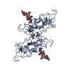
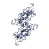
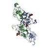
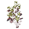
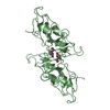
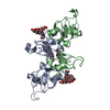
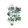
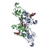
| Title | 2.2 ANGSTROMS RESOLUTION STRUCTURE ANALYSIS OF TWO REFINED N-ACETYLNEURAMINYLLACTOSE-WHEAT GERM AGGLUTININ ISOLECTIN COMPLEXES | ||||||||||||
 Components Components | WHEAT GERM LECTIN | ||||||||||||
 Keywords Keywords | LECTIN (AGGLUTININ) | ||||||||||||
| Function / homology |  Function and homology information Function and homology information | ||||||||||||
| Biological species |   Triticum aestivum (bread wheat) Triticum aestivum (bread wheat) | ||||||||||||
| Method | X-RAY DIFFRACTION / Resolution: 1.8 Å | ||||||||||||
 Authors Authors | Wright, C.S. | ||||||||||||
 Citation Citation | Journal: J.Mol.Biol. / Year: 1990 Title: 2.2 A resolution structure analysis of two refined N-acetylneuraminyl-lactose--wheat germ agglutinin isolectin complexes. Authors: Wright, C.S. #1: Journal: J.Mol.Biol. / Year: 1989Title: Comparison of the Refined Crystal Structures of Two Wheat Germ Isolectins Authors: Wright, C.S. #2: Journal: J.Mol.Evol. / Year: 1989Title: Sequence Variability in Three Wheat Germ Agglutinin Isolectins. Products of Multiple Genes in Polyploid Wheat Authors: Wright, C.S. / Raikhel, N. #3: Journal: J.Mol.Biol. / Year: 1988Title: Erratum. Refinement of the Crystal Structure of Wheat Germ Agglutinin Isolectin 2 at 1.8 Angstroms Resolution Authors: Wright, C.S. #4: Journal: J.Mol.Biol. / Year: 1987Title: Refinement of the Crystal Structure of Wheat Germ Agglutinin Isolectin 2 at 1.8 Angstroms Resolution Authors: Wright, C.S. #5: Journal: J.Mol.Biol. / Year: 1987Title: Preliminary X-Ray Diffraction Results on Co-Crystals of Wheat Germ Agglutinin with a Sialoglycopeptide from the Red Cell Receptor Glycophorina Authors: Wright, C.S. / Kahane, I. #6: Journal: J.Biol.Chem. / Year: 1986Title: Structural Differences in the Two Major Wheat Germ Agglutinin Isolectins Authors: Wright, C.S. / Olafsdottir, S. #7: Journal: J.Mol.Evol. / Year: 1985Title: Evolution of the Multidomain Protein Wheat Germ Agglutinin Authors: Wright, H.T. / Brooks, D.M. / Wright, C.S. #8: Journal: J.Mol.Biol. / Year: 1984Title: Structural Comparison of the Two Distinct Sugar Binding Sites in Wheat Germ Agglutinin Isolectin II Authors: Wright, C.S. #9: Journal: Biochemistry / Year: 1984Title: Primary Structure of Wheat Germ Agglutinin Isolectin 2. Peptide Order Deduced from X-Ray Structure Authors: Wright, C.S. / Gavilanes, F. / Peterson, D.L. #10: Journal: J.Mol.Biol. / Year: 1981Title: Histidine Determination in Wheat Germ Agglutinin Isolectin by X-Ray Diffraction Analysis Authors: Wright, C.S. #11: Journal: Biomolecular Structure, Conformation, Function and EvolutionYear: 1980 Title: Multi-Domain Structure of the Dimeric Lectin Wheat Germ Agglutinin Authors: Wright, C.S. #12: Journal: J.Mol.Biol. / Year: 1980Title: Location of the N-Acetyl-D-Neuraminic Acid Binding Site in Wheat Germ Agglutinin. A Crystallographic Study at 2.8 Angstroms Resolution Authors: Wright, C.S. #13: Journal: J.Biol.Chem. / Year: 1980Title: The Toxin-Agglutinin Fold. A New Group of Small Protein Structures Organized Around a Four-Disulfide Core Authors: Drenth, J. / Low, B.W. / Richardson, J.S. / Wright, C.S. #14: Journal: J.Mol.Biol. / Year: 1980Title: Crystallographic Elucidation of the Saccharide Binding Mode in Wheat Germ Agglutinin and its Biological Significance Authors: Wright, C.S. #15: Journal: J.Mol.Biol. / Year: 1977Title: The Crystal Structure of Wheat Germ Agglutinin at 2.2 Angstroms Resolution Authors: Wright, C.S. #16: Journal: J.Mol.Biol. / Year: 1974Title: Non-Crystallographic Symmetry in the Crystal Dimer of Wheat Germ Agglutinin Authors: Wright, C.S. #17: Journal: J.Mol.Biol. / Year: 1974Title: A Preliminary Crystallographic Study of Wheat Germ Agglutinin Authors: Wright, C.S. / Keith, C. / Langridge, R. / Nagata, Y. / Burger, M.M. | ||||||||||||
| History |
|
- Structure visualization
| Structure viewer | Molecule: MolmilJmol/JSmol |
|---|
- Downloads & links
-Download
| PDBx/mmCIF format | 9wga.cif.gz | 72.2 KB | Display | PDBx/mmCIF format |
|---|---|---|---|---|
| PDB format | pdb9wga.ent.gz | 58.2 KB | Display | PDB format |
| PDBx/mmJSON format | 9wga.json.gz | Tree view | PDBx/mmJSON format | |
| Others |  Other downloads Other downloads |
-Validation report
| Arichive directory | https://data.pdbj.org/pub/pdb/validation_reports/wg/9wgaftp://data.pdbj.org/pub/pdb/validation_reports/wg/9wga | HTTPS FTP |
|---|
-Related structure data


-Links
 PDBj
PDBj- Assembly
| Deposited unit | 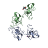
| ||||||||||||||||||||||||
|---|---|---|---|---|---|---|---|---|---|---|---|---|---|---|---|---|---|---|---|---|---|---|---|---|---|
| 1 | 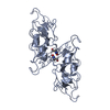
| ||||||||||||||||||||||||
| 2 | 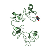
| ||||||||||||||||||||||||
| 3 | 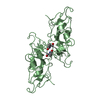
| ||||||||||||||||||||||||
| Unit cell |
| ||||||||||||||||||||||||
| Components on special symmetry positions |
| ||||||||||||||||||||||||
| Noncrystallographic symmetry (NCS) | NCS oper: (Code: given / Matrix: (1),: | Details | THE TRANSFORMATION GIVEN ON THE *MTRIX* RECORDS BELOW YIELDS COORDINATES FOR PROTOMER II WHEN APPLIED TO PROTOMER I. IT REPRESENTS THE NON-CRYSTALLOGRAPHIC SCREW DIAD AXIS RELATING PROTOMERS OF ADJACENT DIMERS. | |
-Components
| #1: Protein | Mass: 17135.109 Da / Num. of mol.: 2 Source method: isolated from a genetically manipulated source Source: (gene. exp.) Triticum aestivum (bread wheat) / References: UniProt: P02876#2: Water | ChemComp-HOH / | Water Mass: 18.015 Da / Num. of mol.: 231 / Source method: isolated from a natural source / Formula: H2O Mass: 18.015 Da / Num. of mol.: 231 / Source method: isolated from a natural source / Formula: H2OCompound details | ALPHA-HELIX AND BETA-SHEET ARE VIRTUALLY ABSENT IN THE STRUCTURAL | |
|---|
-Experimental details
-Experiment
| Experiment | Method: X-RAY DIFFRACTION |
|---|
- Sample preparation
| Crystal | Density Matthews: 2.5 Å3/Da / Density % sol: 50.76 % | ||||||||||||||||||||||||
|---|---|---|---|---|---|---|---|---|---|---|---|---|---|---|---|---|---|---|---|---|---|---|---|---|---|
| Crystal grow | *PLUS Temperature: 37 ℃ / pH: 4.9 / Method: unknown / Details: pH was adjusted to 4.9 with NaOH | ||||||||||||||||||||||||
| Components of the solutions | *PLUS
|
- Processing
| Software | Name: PROLSQ / Classification: refinement | ||||||||||||||||||||||||||||||||||||||||||||||||||||||||||||||||||||||||||||||||||||
|---|---|---|---|---|---|---|---|---|---|---|---|---|---|---|---|---|---|---|---|---|---|---|---|---|---|---|---|---|---|---|---|---|---|---|---|---|---|---|---|---|---|---|---|---|---|---|---|---|---|---|---|---|---|---|---|---|---|---|---|---|---|---|---|---|---|---|---|---|---|---|---|---|---|---|---|---|---|---|---|---|---|---|---|---|---|
| Refinement | Resolution: 1.8→8 Å / σ(F): 3 /
| ||||||||||||||||||||||||||||||||||||||||||||||||||||||||||||||||||||||||||||||||||||
| Refinement step | Cycle: LAST / Resolution: 1.8→8 Å
| ||||||||||||||||||||||||||||||||||||||||||||||||||||||||||||||||||||||||||||||||||||
| Refine LS restraints |
| ||||||||||||||||||||||||||||||||||||||||||||||||||||||||||||||||||||||||||||||||||||
| Refinement | *PLUS Highest resolution: 1.8 Å / Lowest resolution: 8 Å / Num. reflection obs: 17318 / σ(F): 3 / Rfactor obs: 0.175 | ||||||||||||||||||||||||||||||||||||||||||||||||||||||||||||||||||||||||||||||||||||
| Solvent computation | *PLUS | ||||||||||||||||||||||||||||||||||||||||||||||||||||||||||||||||||||||||||||||||||||
| Displacement parameters | *PLUS |
