 Movie
Movie Controller

[English] 日本語
 Yorodumi
- PDB-9rsa: CRYSTAL STRUCTURE OF TWO COVALENT NUCLEOSIDE DERIVATIVES OF RIBON... -
+ Open data

- Basic information
| Entry | Database: PDB / ID: 9rsa | ||||||
|---|---|---|---|---|---|---|---|
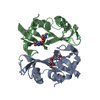
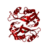
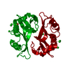
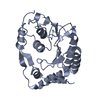
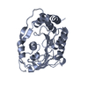
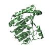
| Title | CRYSTAL STRUCTURE OF TWO COVALENT NUCLEOSIDE DERIVATIVES OF RIBONUCLEASE A | ||||||
 Components Components | RIBONUCLEASE A Pancreatic ribonuclease family Pancreatic ribonuclease family | ||||||
 Keywords Keywords | HYDROLASE (PHOSPHORIC DIESTER) | ||||||
| Function / homology |  Function and homology informationpancreatic ribonuclease / ribonuclease A activity / RNA nuclease activity / nucleic acid binding / lyase activity / defense response to Gram-positive bacterium / extracellular region Function and homology informationpancreatic ribonuclease / ribonuclease A activity / RNA nuclease activity / nucleic acid binding / lyase activity / defense response to Gram-positive bacterium / extracellular regionSimilarity search - Function | ||||||
| Biological species |  Bos taurus (cattle) Bos taurus (cattle) | ||||||
| Method | X-RAY DIFFRACTION / Resolution: 1.8 Å | ||||||
 Authors Authors | Nachman, J. / Wlodawer, A. | ||||||
 Citation Citation | Journal: Biochemistry / Year: 1990 Title: Crystal structure of two covalent nucleoside derivatives of ribonuclease A. Authors: Nachman, J. / Miller, M. / Gilliland, G.L. / Carty, R. / Pincus, M. / Wlodawer, A. #1: Journal: Biochemistry / Year: 1988Title: Structure of Phosphate-Free Ribonuclease A Refined at 1.26 Angstroms Authors: Wlodawer, A. / Svensson, L.A. / Sjolin, L. / Gilliland, G.L. #2: Journal: Proteins / Year: 1986Title: Multiple Conformations of Amino Acid Residues in Ribonuclease A Authors: Svensson, L.A. / Sjolin, L. / Gilliland, G.L. / Finzel, B.C. / Wlodawer, A. #3: Journal: Acta Crystallogr.,Sect.B / Year: 1986Title: Comparison of Two Independently Refined Models of Ribonuclease-A Authors: Wlodawer, A. / Borkakoti, N. / Moss, D.S. / Howlin, B. #4: Journal: J.Biol.Chem. / Year: 1982Title: The Refined Crystal Structure of Ribonuclease A at 2.0 Angstroms Resolution Authors: Wlodawer, A. / Bott, R. / Sjolin, L. | ||||||
| History |
| ||||||
| Remark 700 | SHEET THIS STRUCTURE CONTAINS TWO SHEETS. SHEET S1 COMPRISES THREE STRANDS. IN THE SECOND STRAND OF ...SHEET THIS STRUCTURE CONTAINS TWO SHEETS. SHEET S1 COMPRISES THREE STRANDS. IN THE SECOND STRAND OF SHEET S1, RESIDUES 88 AND 89 *BULGE OUT*. IN ORDER TO REPRESENT THIS BREAK IN STRAND 2, TWO SHEETS (S1A AND S1B) ARE DEFINED BELOW. STRANDS 1 AND 3 OF *SHEETS* S1A AND S1B ARE, THEREFORE, IDENTICAL AND STRAND 2 DIFFERS. SHEET S2 COMPRISES FOUR STRANDS. RESIDUE 120 DOES NOT PROPERLY BELONG IN STRAND 4 OF SHEET S2. IN ORDER TO REPRESENT THIS BREAK IN STRAND 4, TWO SHEETS (S2A AND S2B) ARE DEFINED BELOW. STRANDS 1,2,3 OF *SHEETS* S2A AND S2B ARE, THEREFORE, IDENTICAL AND STRAND 4 DIFFERS. |
- Structure visualization
| Structure viewer | Molecule: MolmilJmol/JSmol |
|---|
- Downloads & links
-Download
| PDBx/mmCIF format | 9rsa.cif.gz | 59.6 KB | Display | PDBx/mmCIF format |
|---|---|---|---|---|
| PDB format | pdb9rsa.ent.gz | 48.7 KB | Display | PDB format |
| PDBx/mmJSON format | 9rsa.json.gz | Tree view | PDBx/mmJSON format | |
| Others |  Other downloads Other downloads |
-Validation report
| Arichive directory | https://data.pdbj.org/pub/pdb/validation_reports/rs/9rsaftp://data.pdbj.org/pub/pdb/validation_reports/rs/9rsa | HTTPS FTP |
|---|
-Related structure data


-Links
 PDBj
PDBj

- Assembly
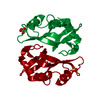
| Deposited unit | 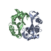
| ||||||||
|---|---|---|---|---|---|---|---|---|---|
| 1 |
| ||||||||
| Unit cell |
| ||||||||
| Atom site foot note | 1: RESIDUES 93 AND 114 OF EACH CHAIN ARE CIS PROLINES. / 2: IN ADU A 125 ONLY THE AMINOACYL LINK IS PRESENT. / 3: SEE REMARK 9. |
-Components
| #1: Protein | Pancreatic ribonuclease family Mass: 13708.326 Da / Num. of mol.: 2 Source method: isolated from a genetically manipulated source Source: (gene. exp.) Bos taurus (cattle) / Cell line: S2 / Organ: PANCREAS / References: UniProt: P61823, EC: 3.1.27.5#2: Chemical |   Mass: 285.253 Da / Num. of mol.: 2 / Source method: obtained synthetically / Formula: C11H15N3O6 Mass: 285.253 Da / Num. of mol.: 2 / Source method: obtained synthetically / Formula: C11H15N3O6#3: Water | ChemComp-HOH / | Water Mass: 18.015 Da / Num. of mol.: 181 / Source method: isolated from a natural source / Formula: H2O Mass: 18.015 Da / Num. of mol.: 181 / Source method: isolated from a natural source / Formula: H2O |
|---|
-Experimental details
-Experiment
| Experiment | Method: X-RAY DIFFRACTION |
|---|
- Sample preparation
| Crystal | Density Matthews: 2.25 Å3/Da / Density % sol: 45.45 % | ||||||||||||||||||||||||
|---|---|---|---|---|---|---|---|---|---|---|---|---|---|---|---|---|---|---|---|---|---|---|---|---|---|
| Crystal grow | *PLUS pH: 5.1 / Method: unknown / Details: pH was adjusted to 5.1 with acetate buffer | ||||||||||||||||||||||||
| Components of the solutions | *PLUS
|
-Data collection
| Reflection | *PLUS Highest resolution: 1.8 Å / Lowest resolution: 2 Å / Num. obs: 19632 / Observed criterion σ(I): 1.5 / Num. measured all: 162247 / Rmerge(I) obs: 0.078 |
|---|
- Processing
| Software | Name: PROFFT / Classification: refinement | ||||||||||||||||||||||||||||||||||||||||||||||||||||||||||||||||||||||||||||||||||||
|---|---|---|---|---|---|---|---|---|---|---|---|---|---|---|---|---|---|---|---|---|---|---|---|---|---|---|---|---|---|---|---|---|---|---|---|---|---|---|---|---|---|---|---|---|---|---|---|---|---|---|---|---|---|---|---|---|---|---|---|---|---|---|---|---|---|---|---|---|---|---|---|---|---|---|---|---|---|---|---|---|---|---|---|---|---|
| Refinement | Resolution: 1.8→10 Å / Rfactor obs: 0.196 | ||||||||||||||||||||||||||||||||||||||||||||||||||||||||||||||||||||||||||||||||||||
| Refinement step | Cycle: LAST / Resolution: 1.8→10 Å
| ||||||||||||||||||||||||||||||||||||||||||||||||||||||||||||||||||||||||||||||||||||
| Refine LS restraints |
| ||||||||||||||||||||||||||||||||||||||||||||||||||||||||||||||||||||||||||||||||||||
| Refinement | *PLUS Highest resolution: 1.8 Å / Lowest resolution: 10 Å / Rfactor obs: 0.196 | ||||||||||||||||||||||||||||||||||||||||||||||||||||||||||||||||||||||||||||||||||||
| Solvent computation | *PLUS | ||||||||||||||||||||||||||||||||||||||||||||||||||||||||||||||||||||||||||||||||||||
| Displacement parameters | *PLUS | ||||||||||||||||||||||||||||||||||||||||||||||||||||||||||||||||||||||||||||||||||||
| Refine LS restraints | *PLUS
|
