 Movie
Movie Controller

[English] 日本語
 Yorodumi
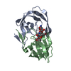
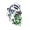
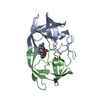
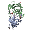
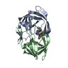
- PDB-8hvp: STRUCTURE AT 2.5-ANGSTROMS RESOLUTION OF CHEMICALLY SYNTHESIZED H... -
+ Open data

- Basic information
| Entry | Database: PDB / ID: 8hvp | |||||||||
|---|---|---|---|---|---|---|---|---|---|---|
| Title | STRUCTURE AT 2.5-ANGSTROMS RESOLUTION OF CHEMICALLY SYNTHESIZED HUMAN IMMUNODEFICIENCY VIRUS TYPE 1 PROTEASE COMPLEXED WITH A HYDROXYETHYLENE*-BASED INHIBITOR | |||||||||
 Components Components |
| |||||||||
 Keywords Keywords | HYDROLASE/HYDROLASE INHIBITOR /  ACID PROTEINASE / HYDROLASE-HYDROLASE INHIBITOR COMPLEX ACID PROTEINASE / HYDROLASE-HYDROLASE INHIBITOR COMPLEX | |||||||||
| Function / homology |  Function and homology informationHIV-1 retropepsin / : / retroviral ribonuclease H / exoribonuclease H / : / exoribonuclease H activity / host multivesicular body / DNA integration / RNA-directed DNA polymerase / viral genome integration into host DNA ...HIV-1 retropepsin / : / retroviral ribonuclease H / exoribonuclease H / : / exoribonuclease H activity / host multivesicular body / DNA integration / RNA-directed DNA polymerase / viral genome integration into host DNA / viral penetration into host nucleus / establishment of integrated proviral latency / RNA-directed DNA polymerase activity / Transferases; Transferring phosphorus-containing groups; Nucleotidyltransferases / RNA-DNA hybrid ribonuclease activity / viral nucleocapsid / DNA recombination / Hydrolases; Acting on ester bonds / DNA-directed DNA polymerase / aspartic-type endopeptidase activity / DNA-directed DNA polymerase activity / symbiont entry into host cell / symbiont-mediated suppression of host gene expression / lipid binding / host cell nucleus / structural molecule activity / host cell plasma membrane / virion membrane / proteolysis / DNA binding / RNA binding / zinc ion binding / membrane Function and homology informationHIV-1 retropepsin / : / retroviral ribonuclease H / exoribonuclease H / : / exoribonuclease H activity / host multivesicular body / DNA integration / RNA-directed DNA polymerase / viral genome integration into host DNA ...HIV-1 retropepsin / : / retroviral ribonuclease H / exoribonuclease H / : / exoribonuclease H activity / host multivesicular body / DNA integration / RNA-directed DNA polymerase / viral genome integration into host DNA / viral penetration into host nucleus / establishment of integrated proviral latency / RNA-directed DNA polymerase activity / Transferases; Transferring phosphorus-containing groups; Nucleotidyltransferases / RNA-DNA hybrid ribonuclease activity / viral nucleocapsid / DNA recombination / Hydrolases; Acting on ester bonds / DNA-directed DNA polymerase / aspartic-type endopeptidase activity / DNA-directed DNA polymerase activity / symbiont entry into host cell / symbiont-mediated suppression of host gene expression / lipid binding / host cell nucleus / structural molecule activity / host cell plasma membrane / virion membrane / proteolysis / DNA binding / RNA binding / zinc ion binding / membraneSimilarity search - Function | |||||||||
| Biological species |   Human immunodeficiency virus 1 Human immunodeficiency virus 1 | |||||||||
| Method | X-RAY DIFFRACTION / Resolution: 2.5 Å | |||||||||
 Authors Authors | Jaskolski, M. / Miller, M. / Tomasselli, A.G. / Sawyer, T.K. / Staples, D.G. / Heinrikson, R.L. / Schneider, J. / Kent, S.B.H. / Wlodawer, A. | |||||||||
 Citation Citation | Journal: Biochemistry / Year: 1991 Title: Structure at 2.5-A resolution of chemically synthesized human immunodeficiency virus type 1 protease complexed with a hydroxyethylene-based inhibitor. Authors: Jaskolski, M. / Tomasselli, A.G. / Sawyer, T.K. / Staples, D.G. / Heinrikson, R.L. / Schneider, J. / Kent, S.B. / Wlodawer, A. #1: Journal: Science / Year: 1989Title: Conserved Folding in Retroviral Proteases. Crystal Structure of a Synthetic HIV-1 Protease Authors: Wlodawer, A. / Miller, M. / Jaskolski, M. / Sathyanarayana, B.K. / Baldwin, E. / Weber, I.T. / Selk, L.M. / Clawson, L. / Schneider, J. / Kent, S.B.H. #2: Journal: Science / Year: 1989Title: Molecular Modeling of the HIV-1 Protease and its Substrate Binding Site Authors: Weber, I.T. / Miller, M. / Jaskolski, M. / Leis, J. / Skalka, A.M. / Wlodawer, A. #3: Journal: Nature / Year: 1989Title: Crystal Structure of a Retroviral Protease Proves Relationship to Aspartic Protease Family Authors: Miller, M. / Jaskolski, M. / Rao, J.K.M. / Leis, J. / Wlodawer, A. #4: Journal: Cell(Cambridge,Mass.) / Year: 1988Title: Enzymatic Activity of a Synthetic 99 Residue Protein Corresponding to the Putative HIV-1 Protease Authors: Schneider, J. / Kent, S.B.H. | |||||||||
| History |
| |||||||||
| Remark 700 | SHEET THE DIMER INTERFACE IS COMPOSED OF INTERDIGITATED N- AND C-TERMINI FROM BOTH SUBUNITS FORMING ...SHEET THE DIMER INTERFACE IS COMPOSED OF INTERDIGITATED N- AND C-TERMINI FROM BOTH SUBUNITS FORMING A FOUR-STRANDED ANTIPARALLEL BETA-SHEET. |
- Structure visualization
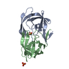
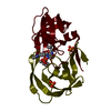
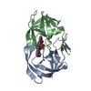
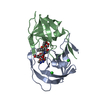
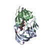
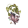
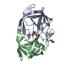
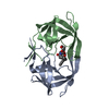
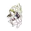
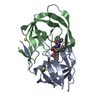
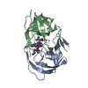
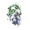
| Structure viewer | Molecule: MolmilJmol/JSmol |
|---|
- Downloads & links
-Download
| PDBx/mmCIF format | 8hvp.cif.gz | 56 KB | Display | PDBx/mmCIF format |
|---|---|---|---|---|
| PDB format | pdb8hvp.ent.gz | 39.1 KB | Display | PDB format |
| PDBx/mmJSON format | 8hvp.json.gz | Tree view | PDBx/mmJSON format | |
| Others |  Other downloads Other downloads |
-Validation report
| Arichive directory | https://data.pdbj.org/pub/pdb/validation_reports/hv/8hvpftp://data.pdbj.org/pub/pdb/validation_reports/hv/8hvp | HTTPS FTP |
|---|
-Related structure data
| Similar structure data |
|---|


-Links
 PDBj
PDBj

- Assembly
| Deposited unit | 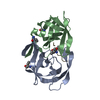
| ||||||||
|---|---|---|---|---|---|---|---|---|---|
| 1 |
| ||||||||
| Unit cell |
| ||||||||
| Atom site foot note | 1: THE INHIBITOR RESIDUE LOV I 5 IS LEU-VAL WITH THE PEPTIDE BOND REPLACED BY CHOH-CH2. |
-Components
| #1: Protein | Mass: 10764.636 Da / Num. of mol.: 2 Source method: isolated from a genetically manipulated source Source: (gene. exp.) Human immunodeficiency virus 1 / Genus: Lentivirus / Production host:  Escherichia coli (E. coli) / References: UniProt: P03369 Escherichia coli (E. coli) / References: UniProt: P03369#2: Protein/peptide | |   Type: Peptide-like / Class: Inhibitor / Mass: 872.061 Da / Num. of mol.: 1 / Source method: obtained synthetically Type: Peptide-like / Class: Inhibitor / Mass: 872.061 Da / Num. of mol.: 1 / Source method: obtained syntheticallyReferences: INHIBITOR VAL-SER-GLN-ASN-LEU-PSI(CH(OH)-CH2)-VAL-ILE-VAL (U-85548E) #3: Water | ChemComp-HOH / | Water Mass: 18.015 Da / Num. of mol.: 80 / Source method: isolated from a natural source / Formula: H2O Mass: 18.015 Da / Num. of mol.: 80 / Source method: isolated from a natural source / Formula: H2ONonpolymer details | THE PEPTIDE BOND BETWEEN RESIDUES LEU I 5 AND VAL I 6 HAS BEEN REPLACED BY A HYDROXYETHYLENE GROUP ...THE PEPTIDE BOND BETWEEN RESIDUES LEU I 5 AND VAL I 6 HAS BEEN REPLACED BY A HYDROXYETH | |
|---|
-Experimental details
-Experiment
| Experiment | Method: X-RAY DIFFRACTION |
|---|
- Sample preparation
| Crystal | Density Matthews: 2.09 Å3/Da / Density % sol: 41.03 % |
|---|---|
| Crystal grow | *PLUS Method: vapor diffusion, hanging drop / Details: referred to Science 246.1149-1152 1989 |
| Components of the solutions | *PLUS Conc.: 60 % / Common name: ammonium sulfate |
-Data collection
| Radiation | Scattering type: x-ray |
|---|---|
| Radiation wavelength | Relative weight: 1 |
| Reflection | *PLUS Highest resolution: 2.5 Å / Num. all: 25199 / Num. obs: 6304 |
- Processing
| Software | Name: PROFFT / Classification: refinement | ||||||||||||||||||||||||||||||||||||||||||||||||||||||||||||||||||||||||||||||||||||
|---|---|---|---|---|---|---|---|---|---|---|---|---|---|---|---|---|---|---|---|---|---|---|---|---|---|---|---|---|---|---|---|---|---|---|---|---|---|---|---|---|---|---|---|---|---|---|---|---|---|---|---|---|---|---|---|---|---|---|---|---|---|---|---|---|---|---|---|---|---|---|---|---|---|---|---|---|---|---|---|---|---|---|---|---|---|
| Refinement | Resolution: 2.5→10 Å /
| ||||||||||||||||||||||||||||||||||||||||||||||||||||||||||||||||||||||||||||||||||||
| Refinement step | Cycle: LAST / Resolution: 2.5→10 Å
| ||||||||||||||||||||||||||||||||||||||||||||||||||||||||||||||||||||||||||||||||||||
| Refine LS restraints |
| ||||||||||||||||||||||||||||||||||||||||||||||||||||||||||||||||||||||||||||||||||||
| Refinement | *PLUS Highest resolution: 2.5 Å / Lowest resolution: 10 Å / Num. reflection obs: 4768 / Rfactor obs: 0.138 | ||||||||||||||||||||||||||||||||||||||||||||||||||||||||||||||||||||||||||||||||||||
| Solvent computation | *PLUS | ||||||||||||||||||||||||||||||||||||||||||||||||||||||||||||||||||||||||||||||||||||
| Displacement parameters | *PLUS |
