 Movie
Movie Controller

[English] 日本語
 Yorodumi
- PDB-7jx3: Mapping neutralizing and immunodominant sites on the SARS-CoV-2 s... -
+ Open data

- Basic information
| Entry | Database: PDB / ID: 7jx3 | |||||||||
|---|---|---|---|---|---|---|---|---|---|---|
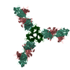
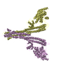
| Title | Mapping neutralizing and immunodominant sites on the SARS-CoV-2 spike receptor-binding domain by structure-guided high-resolution serology | |||||||||
 Components Components |
| |||||||||
 Keywords Keywords |  VIRAL PROTEIN/IMMUNE SYSTEM / COVID-19 / SARS-CoV-2 / neutralizing monoclonal antibody / VIRAL PROTEIN-IMMUNE SYSTEM complex VIRAL PROTEIN/IMMUNE SYSTEM / COVID-19 / SARS-CoV-2 / neutralizing monoclonal antibody / VIRAL PROTEIN-IMMUNE SYSTEM complex | |||||||||
| Function / homology |  Function and homology information Function and homology informationMaturation of spike protein / viral translation / Translation of Structural Proteins / Virion Assembly and Release / host cell surface / host extracellular space / suppression by virus of host tetherin activity / Induction of Cell-Cell Fusion / structural constituent of virion / host cell endoplasmic reticulum-Golgi intermediate compartment membrane ...Maturation of spike protein / viral translation / Translation of Structural Proteins / Virion Assembly and Release / host cell surface / host extracellular space / suppression by virus of host tetherin activity / Induction of Cell-Cell Fusion / structural constituent of virion / host cell endoplasmic reticulum-Golgi intermediate compartment membrane / entry receptor-mediated virion attachment to host cell / receptor-mediated endocytosis of virus by host cell / Attachment and Entry / membrane fusion / positive regulation of viral entry into host cell / receptor-mediated virion attachment to host cell / receptor ligand activity / host cell surface receptor binding / fusion of virus membrane with host plasma membrane / fusion of virus membrane with host endosome membrane / viral envelope / symbiont-mediated suppression of host type I interferon-mediated signaling pathway / virion attachment to host cell / SARS-CoV-2 activates/modulates innate and adaptive immune responses / host cell plasma membrane / virion membrane / membrane / identical protein binding / plasma membraneSimilarity search - Function | |||||||||
| Biological species |  Homo sapiens (human) Homo sapiens (human)  Severe acute respiratory syndrome coronavirus 2 Severe acute respiratory syndrome coronavirus 2 | |||||||||
| Method | X-RAY DIFFRACTION / SYNCHROTRON / MOLECULAR REPLACEMENT / molecular replacement / Resolution: 2.65 Å | |||||||||
 Authors Authors | Snell, G. / Czudnochowski, N. / Rosen, L.E. / Nix, J.C. / Corti, D. / Veesler, D. / Park, Y.J. / Walls, A.C. / Tortorici, M.A. / Cameroni, E. ...Snell, G. / Czudnochowski, N. / Rosen, L.E. / Nix, J.C. / Corti, D. / Veesler, D. / Park, Y.J. / Walls, A.C. / Tortorici, M.A. / Cameroni, E. / Pinto, D. / Beltramello, M. / Seattle Structural Genomics Center for Infectious Disease (SSGCID) | |||||||||
| Funding support |  United States, 2items United States, 2items
| |||||||||
 Citation Citation | Journal: Cell / Year: 2020 Title: Mapping Neutralizing and Immunodominant Sites on the SARS-CoV-2 Spike Receptor-Binding Domain by Structure-Guided High-Resolution Serology. Authors: Luca Piccoli / Young-Jun Park / M Alejandra Tortorici / Nadine Czudnochowski / Alexandra C Walls / Martina Beltramello / Chiara Silacci-Fregni / Dora Pinto / Laura E Rosen / John E Bowen / ...Authors: Luca Piccoli / Young-Jun Park / M Alejandra Tortorici / Nadine Czudnochowski / Alexandra C Walls / Martina Beltramello / Chiara Silacci-Fregni / Dora Pinto / Laura E Rosen / John E Bowen / Oliver J Acton / Stefano Jaconi / Barbara Guarino / Andrea Minola / Fabrizia Zatta / Nicole Sprugasci / Jessica Bassi / Alessia Peter / Anna De Marco / Jay C Nix / Federico Mele / Sandra Jovic / Blanca Fernandez Rodriguez / Sneha V Gupta / Feng Jin / Giovanni Piumatti / Giorgia Lo Presti / Alessandra Franzetti Pellanda / Maira Biggiogero / Maciej Tarkowski / Matteo S Pizzuto / Elisabetta Cameroni / Colin Havenar-Daughton / Megan Smithey / David Hong / Valentino Lepori / Emiliano Albanese / Alessandro Ceschi / Enos Bernasconi / Luigia Elzi / Paolo Ferrari / Christian Garzoni / Agostino Riva / Gyorgy Snell / Federica Sallusto / Katja Fink / Herbert W Virgin / Antonio Lanzavecchia / Davide Corti / David Veesler /     Abstract: Analysis of the specificity and kinetics of neutralizing antibodies (nAbs) elicited by SARS-CoV-2 infection is crucial for understanding immune protection and identifying targets for vaccine design. ...Analysis of the specificity and kinetics of neutralizing antibodies (nAbs) elicited by SARS-CoV-2 infection is crucial for understanding immune protection and identifying targets for vaccine design. In a cohort of 647 SARS-CoV-2-infected subjects, we found that both the magnitude of Ab responses to SARS-CoV-2 spike (S) and nucleoprotein and nAb titers correlate with clinical scores. The receptor-binding domain (RBD) is immunodominant and the target of 90% of the neutralizing activity present in SARS-CoV-2 immune sera. Whereas overall RBD-specific serum IgG titers waned with a half-life of 49 days, nAb titers and avidity increased over time for some individuals, consistent with affinity maturation. We structurally defined an RBD antigenic map and serologically quantified serum Abs specific for distinct RBD epitopes leading to the identification of two major receptor-binding motif antigenic sites. Our results explain the immunodominance of the receptor-binding motif and will guide the design of COVID-19 vaccines and therapeutics. | |||||||||
| History |
|
- Structure visualization
| Structure viewer | Molecule: MolmilJmol/JSmol |
|---|
- Downloads & links
-Download
| PDBx/mmCIF format | 7jx3.cif.gz | 589.8 KB | Display | PDBx/mmCIF format |
|---|---|---|---|---|
| PDB format | pdb7jx3.ent.gz | 487.9 KB | Display | PDB format |
| PDBx/mmJSON format | 7jx3.json.gz | Tree view | PDBx/mmJSON format | |
| Others |  Other downloads Other downloads |
-Validation report
| Arichive directory | https://data.pdbj.org/pub/pdb/validation_reports/jx/7jx3ftp://data.pdbj.org/pub/pdb/validation_reports/jx/7jx3 | HTTPS FTP |
|---|
-Related structure data
| Related structure data |  7jv2C  7jv4C  7jv6C  7jvaC  7jvcC  7jw0C 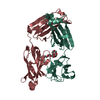 7jxcC 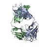 7jxdC 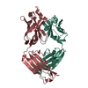 7jxeC 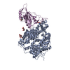 6m0jS S: Starting model for refinement C: citing same article ( |
|---|---|
| Similar structure data |


-Links
 PDBj
PDBj


- Assembly
| Deposited unit | 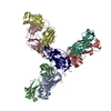
| ||||||||
|---|---|---|---|---|---|---|---|---|---|
| 1 |
| ||||||||
| Unit cell |
|
-Components
-Antibody , 6 types, 6 molecules BADCLH
| #1: Antibody | Mass: 23204.697 Da / Num. of mol.: 1 Source method: isolated from a genetically manipulated source Source: (gene. exp.) Homo sapiens (human) / Production host: Homo sapiens (human) |
|---|---|
| #2: Antibody | Mass: 24573.471 Da / Num. of mol.: 1 Source method: isolated from a genetically manipulated source Source: (gene. exp.) Homo sapiens (human) / Production host: Homo sapiens (human) |
| #3: Antibody | Mass: 23139.297 Da / Num. of mol.: 1 Source method: isolated from a genetically manipulated source Source: (gene. exp.) Homo sapiens (human) / Production host: Homo sapiens (human) |
| #4: Antibody | Mass: 24246.059 Da / Num. of mol.: 1 Source method: isolated from a genetically manipulated source Source: (gene. exp.) Homo sapiens (human) / Production host: Homo sapiens (human) |
| #5: Antibody | Mass: 23370.932 Da / Num. of mol.: 1 Source method: isolated from a genetically manipulated source Source: (gene. exp.) Homo sapiens (human) / Production host: Homo sapiens (human) |
| #6: Antibody | Mass: 23729.389 Da / Num. of mol.: 1 Source method: isolated from a genetically manipulated source Source: (gene. exp.) Homo sapiens (human) / Production host: Homo sapiens (human) |
-Protein / Sugars / Non-polymers , 3 types, 27 molecules R

| #7: Protein | Mass: 22948.783 Da / Num. of mol.: 1 / Fragment: Receptor binding domain (UNP residues 328-531) Source method: isolated from a genetically manipulated source Source: (gene. exp.) Severe acute respiratory syndrome coronavirus 2Gene: S, 2 / Production host: Homo sapiens (human) / References: UniProt: P0DTC2 |
|---|---|
| #8: Sugar | ChemComp-NAG / N-Acetylglucosamine Type: D-saccharide, beta linking / Mass: 221.208 Da / Num. of mol.: 1 / Source method: obtained synthetically / Formula: C8H15NO6 Type: D-saccharide, beta linking / Mass: 221.208 Da / Num. of mol.: 1 / Source method: obtained synthetically / Formula: C8H15NO6 |
| #9: Water | ChemComp-HOH / WaterMass: 18.015 Da / Num. of mol.: 25 / Source method: isolated from a natural source / Formula: H2O |
-Details
| Has ligand of interest | N |
|---|
-Experimental details
-Experiment
| Experiment | Method: X-RAY DIFFRACTION / Number of used crystals: 1 |
|---|
- Sample preparation
| Crystal | Density Matthews: 2.94 Å3/Da / Density % sol: 58.12 % |
|---|---|
| Crystal grow | Temperature: 295 K / Method: vapor diffusion, sitting drop Details: 16.2% w/v PEG4000, 0.09 M sodium citrate, pH 6.0, 0.18 M ammonium acetate, 0.02 M potassium acetate, 0.01 MES, pH 6, 1.5% v/v pentaerythritol ethoxylate (15/4 EO/OH) |
-Data collection
| Diffraction | Mean temperature: 100 K / Serial crystal experiment: N | ||||||||||||||||||||||||||||||
|---|---|---|---|---|---|---|---|---|---|---|---|---|---|---|---|---|---|---|---|---|---|---|---|---|---|---|---|---|---|---|---|
| Diffraction source | Source: SYNCHROTRON / Site: ALS / Beamline: 4.2.2 / Wavelength: 0.97625 Å | ||||||||||||||||||||||||||||||
| Detector | Type: RDI CMOS_8M / Detector: CMOS / Date: Aug 7, 2020 | ||||||||||||||||||||||||||||||
| Radiation | Monochromator: double crystal SI(111) / Protocol: SINGLE WAVELENGTH / Monochromatic (M) / Laue (L): M / Scattering type: x-ray | ||||||||||||||||||||||||||||||
| Radiation wavelength | Wavelength: 0.97625 Å / Relative weight: 1 | ||||||||||||||||||||||||||||||
| Reflection | Resolution: 2.65→47.75 Å / Num. obs: 55237 / % possible obs: 99.6 % / Redundancy: 7.4 % / CC1/2: 0.996 / Rmerge(I) obs: 0.199 / Rpim(I) all: 0.079 / Rrim(I) all: 0.215 / Net I/σ(I): 8.7 / Num. measured all: 406155 | ||||||||||||||||||||||||||||||
| Reflection shell | Diffraction-ID: 1
|
-Phasing
| Phasing | Method: molecular replacement |
|---|
- Processing
| Software |
| ||||||||||||||||||||||||||||||||||||||||||||||||||||||||||||||||||||||||||||||||||||||||||||||||||||||||||||||||||||||||||||||||||||||||||||||||||||||||||||||||||||||||||||||||||||||||||||||||||||||||
|---|---|---|---|---|---|---|---|---|---|---|---|---|---|---|---|---|---|---|---|---|---|---|---|---|---|---|---|---|---|---|---|---|---|---|---|---|---|---|---|---|---|---|---|---|---|---|---|---|---|---|---|---|---|---|---|---|---|---|---|---|---|---|---|---|---|---|---|---|---|---|---|---|---|---|---|---|---|---|---|---|---|---|---|---|---|---|---|---|---|---|---|---|---|---|---|---|---|---|---|---|---|---|---|---|---|---|---|---|---|---|---|---|---|---|---|---|---|---|---|---|---|---|---|---|---|---|---|---|---|---|---|---|---|---|---|---|---|---|---|---|---|---|---|---|---|---|---|---|---|---|---|---|---|---|---|---|---|---|---|---|---|---|---|---|---|---|---|---|---|---|---|---|---|---|---|---|---|---|---|---|---|---|---|---|---|---|---|---|---|---|---|---|---|---|---|---|---|---|---|---|---|
| Refinement | Method to determine structure: MOLECULAR REPLACEMENT Starting model: PDB entry 6M0J Resolution: 2.65→47.5 Å / Cor.coef. Fo:Fc: 0.919 / Cor.coef. Fo:Fc free: 0.899 / SU B: 53.058 / SU ML: 0.439 / SU R Cruickshank DPI: 0.9723 / Cross valid method: THROUGHOUT / σ(F): 0 / ESU R: 0.972 / ESU R Free: 0.367 / Stereochemistry target values: MAXIMUM LIKELIHOOD Details: HYDROGENS HAVE BEEN ADDED IN THE RIDING POSITIONS U VALUES : WITH TLS ADDED
| ||||||||||||||||||||||||||||||||||||||||||||||||||||||||||||||||||||||||||||||||||||||||||||||||||||||||||||||||||||||||||||||||||||||||||||||||||||||||||||||||||||||||||||||||||||||||||||||||||||||||
| Solvent computation | Ion probe radii: 0.8 Å / Shrinkage radii: 0.8 Å / VDW probe radii: 1.2 Å / Solvent model: MASK | ||||||||||||||||||||||||||||||||||||||||||||||||||||||||||||||||||||||||||||||||||||||||||||||||||||||||||||||||||||||||||||||||||||||||||||||||||||||||||||||||||||||||||||||||||||||||||||||||||||||||
| Displacement parameters | Biso max: 166.18 Å2 / Biso mean: 66.749 Å2 / Biso min: 28.4 Å2
| ||||||||||||||||||||||||||||||||||||||||||||||||||||||||||||||||||||||||||||||||||||||||||||||||||||||||||||||||||||||||||||||||||||||||||||||||||||||||||||||||||||||||||||||||||||||||||||||||||||||||
| Refinement step | Cycle: final / Resolution: 2.65→47.5 Å
| ||||||||||||||||||||||||||||||||||||||||||||||||||||||||||||||||||||||||||||||||||||||||||||||||||||||||||||||||||||||||||||||||||||||||||||||||||||||||||||||||||||||||||||||||||||||||||||||||||||||||
| Refine LS restraints |
| ||||||||||||||||||||||||||||||||||||||||||||||||||||||||||||||||||||||||||||||||||||||||||||||||||||||||||||||||||||||||||||||||||||||||||||||||||||||||||||||||||||||||||||||||||||||||||||||||||||||||
| LS refinement shell | Resolution: 2.65→2.719 Å / Rfactor Rfree error: 0 / Total num. of bins used: 20
| ||||||||||||||||||||||||||||||||||||||||||||||||||||||||||||||||||||||||||||||||||||||||||||||||||||||||||||||||||||||||||||||||||||||||||||||||||||||||||||||||||||||||||||||||||||||||||||||||||||||||
| Refinement TLS params. | Method: refined / Refine-ID: X-RAY DIFFRACTION
| ||||||||||||||||||||||||||||||||||||||||||||||||||||||||||||||||||||||||||||||||||||||||||||||||||||||||||||||||||||||||||||||||||||||||||||||||||||||||||||||||||||||||||||||||||||||||||||||||||||||||
| Refinement TLS group |
|
