 Movie
Movie Controller


+ Open data

- Basic information
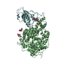
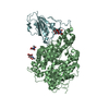
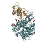
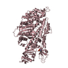
| Entry | Database: PDB / ID: 7c8d | ||||||
|---|---|---|---|---|---|---|---|
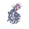
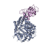
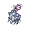
| Title | Cryo-EM structure of cat ACE2 and SARS-CoV-2 RBD | ||||||
 Components Components |
| ||||||
 Keywords Keywords | HYDROLASE/VIRAL PROTEIN /  ACE2 / SARS-CoV-2 / Cryo-EM / complex / PROTEIN BINDING / HYDROLASE-VIRAL PROTEIN complex ACE2 / SARS-CoV-2 / Cryo-EM / complex / PROTEIN BINDING / HYDROLASE-VIRAL PROTEIN complex | ||||||
| Function / homology |  Function and homology informationangiotensin-converting enzyme 2 / Hydrolases; Acting on peptide bonds (peptidases); Metallocarboxypeptidases / peptidyl-dipeptidase activity / carboxypeptidase activity / metallopeptidase activity / virus receptor activity / Maturation of spike protein / viral translation / Translation of Structural Proteins / Virion Assembly and Release ...angiotensin-converting enzyme 2 / Hydrolases; Acting on peptide bonds (peptidases); Metallocarboxypeptidases / peptidyl-dipeptidase activity / carboxypeptidase activity / metallopeptidase activity / virus receptor activity / Maturation of spike protein / viral translation / Translation of Structural Proteins / Virion Assembly and Release / host cell surface / host extracellular space / suppression by virus of host tetherin activity / Induction of Cell-Cell Fusion / structural constituent of virion / host cell endoplasmic reticulum-Golgi intermediate compartment membrane / entry receptor-mediated virion attachment to host cell / receptor-mediated endocytosis of virus by host cell / Attachment and Entry / membrane fusion / positive regulation of viral entry into host cell / receptor-mediated virion attachment to host cell / receptor ligand activity / membrane => GO:0016020 / host cell surface receptor binding / fusion of virus membrane with host plasma membrane / fusion of virus membrane with host endosome membrane / viral envelope / symbiont-mediated suppression of host type I interferon-mediated signaling pathway / virion attachment to host cell / SARS-CoV-2 activates/modulates innate and adaptive immune responses / host cell plasma membrane / virion membrane / cell surface / extracellular space / membrane / identical protein binding / metal ion binding / plasma membrane / cytoplasm Function and homology informationangiotensin-converting enzyme 2 / Hydrolases; Acting on peptide bonds (peptidases); Metallocarboxypeptidases / peptidyl-dipeptidase activity / carboxypeptidase activity / metallopeptidase activity / virus receptor activity / Maturation of spike protein / viral translation / Translation of Structural Proteins / Virion Assembly and Release ...angiotensin-converting enzyme 2 / Hydrolases; Acting on peptide bonds (peptidases); Metallocarboxypeptidases / peptidyl-dipeptidase activity / carboxypeptidase activity / metallopeptidase activity / virus receptor activity / Maturation of spike protein / viral translation / Translation of Structural Proteins / Virion Assembly and Release / host cell surface / host extracellular space / suppression by virus of host tetherin activity / Induction of Cell-Cell Fusion / structural constituent of virion / host cell endoplasmic reticulum-Golgi intermediate compartment membrane / entry receptor-mediated virion attachment to host cell / receptor-mediated endocytosis of virus by host cell / Attachment and Entry / membrane fusion / positive regulation of viral entry into host cell / receptor-mediated virion attachment to host cell / receptor ligand activity / membrane => GO:0016020 / host cell surface receptor binding / fusion of virus membrane with host plasma membrane / fusion of virus membrane with host endosome membrane / viral envelope / symbiont-mediated suppression of host type I interferon-mediated signaling pathway / virion attachment to host cell / SARS-CoV-2 activates/modulates innate and adaptive immune responses / host cell plasma membrane / virion membrane / cell surface / extracellular space / membrane / identical protein binding / metal ion binding / plasma membrane / cytoplasmSimilarity search - Function | ||||||
| Biological species |  Felis catus (domestic cat) Felis catus (domestic cat)  Severe acute respiratory syndrome coronavirus 2 Severe acute respiratory syndrome coronavirus 2 | ||||||
| Method | ELECTRON MICROSCOPY / single particle reconstruction / Resolution: 3 Å | ||||||
 Authors Authors | Gao, G.F. / Wang, Q.H. / Wu, L.l. | ||||||
| Funding support |  China, 1items China, 1items
| ||||||
 Citation Citation | Journal: Cell Discov / Year: 2020 Title: Broad host range of SARS-CoV-2 and the molecular basis for SARS-CoV-2 binding to cat ACE2. Authors: Lili Wu / Qian Chen / Kefang Liu / Jia Wang / Pengcheng Han / Yanfang Zhang / Yu Hu / Yumin Meng / Xiaoqian Pan / Chengpeng Qiao / Siyu Tian / Pei Du / Hao Song / Weifeng Shi / Jianxun Qi / ...Authors: Lili Wu / Qian Chen / Kefang Liu / Jia Wang / Pengcheng Han / Yanfang Zhang / Yu Hu / Yumin Meng / Xiaoqian Pan / Chengpeng Qiao / Siyu Tian / Pei Du / Hao Song / Weifeng Shi / Jianxun Qi / Hong-Wei Wang / Jinghua Yan / George Fu Gao / Qihui Wang /  Abstract: Severe acute respiratory syndrome coronavirus 2 (SARS-CoV-2), the causative agent of the recent pandemic COVID-19, is reported to have originated from bats, with its intermediate host unknown to date. ...Severe acute respiratory syndrome coronavirus 2 (SARS-CoV-2), the causative agent of the recent pandemic COVID-19, is reported to have originated from bats, with its intermediate host unknown to date. Here, we screened 26 animal counterparts of the human ACE2 (hACE2), the receptor for SARS-CoV-2 and SARS-CoV, and found that the ACE2s from various species, including pets, domestic animals and multiple wild animals, could bind to SARS-CoV-2 receptor binding domain (RBD) and facilitate the transduction of SARS-CoV-2 pseudovirus. Comparing to SARS-CoV-2, SARS-CoV seems to have a slightly wider range in choosing its receptor. We further resolved the cryo-electron microscopy (cryo-EM) structure of the cat ACE2 (cACE2) in complex with the SARS-CoV-2 RBD at a resolution of 3 Å, revealing similar binding mode as hACE2 to the SARS-CoV-2 RBD. These results shed light on pursuing the intermediate host of SARS-CoV-2 and highlight the necessity of monitoring susceptible hosts to prevent further outbreaks. | ||||||
| History |
|
- Structure visualization
| Movie |
Movie viewer |
|---|---|
| Structure viewer | Molecule: MolmilJmol/JSmol |
- Downloads & links
-Download
| PDBx/mmCIF format | 7c8d.cif.gz | 202.1 KB | Display | PDBx/mmCIF format |
|---|---|---|---|---|
| PDB format | pdb7c8d.ent.gz | 154.8 KB | Display | PDB format |
| PDBx/mmJSON format | 7c8d.json.gz | Tree view | PDBx/mmJSON format | |
| Others |  Other downloads Other downloads |
-Validation report
| Arichive directory | https://data.pdbj.org/pub/pdb/validation_reports/c8/7c8dftp://data.pdbj.org/pub/pdb/validation_reports/c8/7c8d | HTTPS FTP |
|---|
-Related structure data
| Related structure data |  30305MC M: map data used to model this data C: citing same article ( |
|---|---|
| Similar structure data |


-Links
 PDBj
PDBj


- Assembly
| Deposited unit | 
|
|---|---|
| 1 |
|
-Components
| #1: Protein | / ACE-related carboxypeptidase Mass: 85132.953 Da / Num. of mol.: 1 Source method: isolated from a genetically manipulated source Source: (gene. exp.) Felis catus (domestic cat) / Gene: ACE2 / Production host:  Escherichia coli (E. coli) Escherichia coli (E. coli)References: UniProt: Q56H28, angiotensin-converting enzyme 2 |
|---|---|
| #2: Protein | Mass: 21873.496 Da / Num. of mol.: 1 Source method: isolated from a genetically manipulated source Source: (gene. exp.) Severe acute respiratory syndrome coronavirus 2Gene: S, 2 / Production host:   Spodoptera frugiperda (fall armyworm) / References: UniProt: P0DTC2 Spodoptera frugiperda (fall armyworm) / References: UniProt: P0DTC2 |
| #3: Chemical | ChemComp-ZN /   Mass: 65.409 Da / Num. of mol.: 1 / Source method: obtained synthetically / Formula: Zn / Feature type: SUBJECT OF INVESTIGATION Mass: 65.409 Da / Num. of mol.: 1 / Source method: obtained synthetically / Formula: Zn / Feature type: SUBJECT OF INVESTIGATION |
| Has ligand of interest | Y |
-Experimental details
-Experiment
| Experiment | Method: ELECTRON MICROSCOPY |
|---|---|
| EM experiment | Aggregation state: PARTICLE / 3D reconstruction method: single particle reconstruction |
- Sample preparation
| Component |
| ||||||||||||||||||||||||
|---|---|---|---|---|---|---|---|---|---|---|---|---|---|---|---|---|---|---|---|---|---|---|---|---|---|
| Source (natural) | Organism: Felis catus (domestic cat) | ||||||||||||||||||||||||
| Source (recombinant) | Organism: Escherichia coli (E. coli) | ||||||||||||||||||||||||
| Buffer solution | pH: 8 | ||||||||||||||||||||||||
| Specimen | Embedding applied: NO / Shadowing applied: NO / Staining applied: NO / Vitrification applied: NO |
- Electron microscopy imaging
| Experimental equipment |  Model: Titan Krios / Image courtesy: FEI Company | ||||||||||||
|---|---|---|---|---|---|---|---|---|---|---|---|---|---|
| Microscopy | Model: FEI TITAN KRIOS | ||||||||||||
| Electron gun | Electron source: FIELD EMISSION GUN / Accelerating voltage: 300 kV / Illumination mode: FLOOD BEAM | ||||||||||||
| Electron lens | Mode: BRIGHT FIELDBright-field microscopy | ||||||||||||
| Image recording |
|
- Processing
| Software | Name: PHENIX / Version: 1.17.1_3660: / Classification: refinement | ||||||||||||||||||||||||
|---|---|---|---|---|---|---|---|---|---|---|---|---|---|---|---|---|---|---|---|---|---|---|---|---|---|
| CTF correction | Type: NONE | ||||||||||||||||||||||||
| Symmetry | Point symmetry: C1 (asymmetric) | ||||||||||||||||||||||||
| 3D reconstruction | Resolution: 3 Å / Resolution method: FSC 0.143 CUT-OFF / Num. of particles: 195370 / Symmetry type: POINT | ||||||||||||||||||||||||
| Refine LS restraints |
|
