 Movie
Movie Controller

[English] 日本語
 Yorodumi
- PDB-6y01: The structure of the molybdenum cofactor binding protein from the... -
+ Open data

- Basic information
| Entry | Database: PDB / ID: 6y01 | ||||||
|---|---|---|---|---|---|---|---|
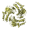
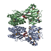
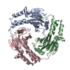
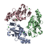
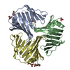
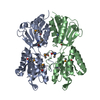
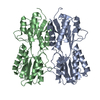
| Title | The structure of the molybdenum cofactor binding protein from the phototrophic bacterium Rippkaea orientalis | ||||||
 Components Components | p450 cytochrome, putative | ||||||
 Keywords Keywords |  TRANSPORT PROTEIN / molybdenum cofactor / Moco / ligand binding / Rossmann fold TRANSPORT PROTEIN / molybdenum cofactor / Moco / ligand binding / Rossmann fold | ||||||
| Function / homology | Conserved hypothetical protein CHP00725 / SLOG cluster4 / SLOG cluster4 family / IMIDAZOLE / p450 cytochrome, putative Function and homology information Function and homology information | ||||||
| Biological species | Rippkaea orientalis | ||||||
| Method | X-RAY DIFFRACTION / SYNCHROTRON / MOLECULAR REPLACEMENT / Resolution: 1.23 Å | ||||||
 Authors Authors | Krausze, J. | ||||||
 Citation Citation | Journal: Acta Crystallogr.,Sect.F / Year: 2020 Title: The structure of the Moco carrier protein from Rippkaea orientalis. Authors: Krausze, J. / Hercher, T.W. / Archna, A. / Kruse, T. | ||||||
| History |
|
- Structure visualization
| Structure viewer | Molecule: MolmilJmol/JSmol |
|---|
- Downloads & links
-Download
| PDBx/mmCIF format | 6y01.cif.gz | 252.7 KB | Display | PDBx/mmCIF format |
|---|---|---|---|---|
| PDB format | pdb6y01.ent.gz | Display | PDB format | |
| PDBx/mmJSON format | 6y01.json.gz | Tree view | PDBx/mmJSON format | |
| Others |  Other downloads Other downloads |
-Validation report
| Arichive directory | https://data.pdbj.org/pub/pdb/validation_reports/y0/6y01ftp://data.pdbj.org/pub/pdb/validation_reports/y0/6y01 | HTTPS FTP |
|---|
-Related structure data
| Related structure data |  2iz6S S: Starting model for refinement |
|---|---|
| Similar structure data |

-Links
 PDBj
PDBj


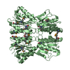
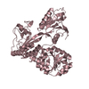
- Assembly
| Deposited unit | 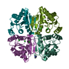
| ||||||||||||||||||||||||||||
|---|---|---|---|---|---|---|---|---|---|---|---|---|---|---|---|---|---|---|---|---|---|---|---|---|---|---|---|---|---|
| 1 |
| ||||||||||||||||||||||||||||
| Unit cell |
| ||||||||||||||||||||||||||||
| Noncrystallographic symmetry (NCS) | NCS domain:
NCS ensembles :
|
-Components
| #1: Protein | Mass: 18893.697 Da / Num. of mol.: 4 Source method: isolated from a genetically manipulated source Source: (gene. exp.)  Rippkaea orientalis (strain PCC 8801) (bacteria) Rippkaea orientalis (strain PCC 8801) (bacteria)Gene: PCC8801_2644 / Plasmid: pQE80 / Cell line (production host): RK5204 / Production host: Escherichia coli (E. coli) / References: UniProt: B7K4Z0#2: Chemical | Imidazole  Mass: 69.085 Da / Num. of mol.: 2 / Source method: obtained synthetically / Formula: C3H5N2 Mass: 69.085 Da / Num. of mol.: 2 / Source method: obtained synthetically / Formula: C3H5N2#3: Chemical | ChemComp-CL / Chloride  Mass: 35.453 Da / Num. of mol.: 10 / Source method: obtained synthetically / Formula: Cl Mass: 35.453 Da / Num. of mol.: 10 / Source method: obtained synthetically / Formula: Cl#4: Water | ChemComp-HOH / | Water Mass: 18.015 Da / Num. of mol.: 393 / Source method: isolated from a natural source / Formula: H2O Mass: 18.015 Da / Num. of mol.: 393 / Source method: isolated from a natural source / Formula: H2OHas ligand of interest | N | |
|---|
-Experimental details
-Experiment
| Experiment | Method: X-RAY DIFFRACTION / Number of used crystals: 1 |
|---|
- Sample preparation
| Crystal | Density Matthews: 2.12 Å3/Da / Density % sol: 41.88 % / Description: rod-like, monoclinic |
|---|---|
| Crystal grow | Temperature: 292 K / Method: vapor diffusion, sitting drop / pH: 6 Details: 15% (v/v) Pentaerythritol ethoxylate; 0.2 M potassium acetate; 3% (v/v) Jeffamine T-403; 0.1 M MES pH 6.0 |
-Data collection
| Diffraction | Mean temperature: 100 K / Serial crystal experiment: N |
|---|---|
| Diffraction source | Source: SYNCHROTRON / Site: SLS  / Beamline: X06DA / Wavelength: 1.00003 Å / Beamline: X06DA / Wavelength: 1.00003 Å |
| Detector | Type: DECTRIS PILATUS 2M-F / Detector: PIXEL / Date: May 17, 2015 / Details: TOROIDAL FOCUSING MIRRORS |
| Radiation | Monochromator: Bartels Monochromator / Protocol: SINGLE WAVELENGTH / Monochromatic (M) / Laue (L): M / Scattering type: x-ray |
| Radiation wavelength | Wavelength: 1.00003 Å / Relative weight: 1 |
| Reflection | Resolution: 1.23→64.52 Å / Num. obs: 181674 / % possible obs: 100 % / Observed criterion σ(I): -3 / Redundancy: 6.7 % / Biso Wilson estimate: 15.66 Å2 / CC1/2: 1 / CC star: 1 / Rmerge(I) obs: 0.05279 / Rpim(I) all: 0.02177 / Rrim(I) all: 0.05719 / Net I/σ(I): 16.35 |
| Reflection shell | Resolution: 1.23→1.274 Å / Redundancy: 6.3 % / Rmerge(I) obs: 3.026 / Mean I/σ(I) obs: 0.55 / Num. unique obs: 18104 / CC1/2: 0.193 / CC star: 0.569 / Rpim(I) all: 1.297 / Rrim(I) all: 3.299 / % possible all: 100 |
- Processing
| Software |
| ||||||||||||||||||||||||||||||||||||||||||||||||||||||||||||||||||||||||||||||||||||||||||||||||||||||||||||||||||||||||||||||||||||||||||||||||||||||||||||||||||||||||||||||||||||||
|---|---|---|---|---|---|---|---|---|---|---|---|---|---|---|---|---|---|---|---|---|---|---|---|---|---|---|---|---|---|---|---|---|---|---|---|---|---|---|---|---|---|---|---|---|---|---|---|---|---|---|---|---|---|---|---|---|---|---|---|---|---|---|---|---|---|---|---|---|---|---|---|---|---|---|---|---|---|---|---|---|---|---|---|---|---|---|---|---|---|---|---|---|---|---|---|---|---|---|---|---|---|---|---|---|---|---|---|---|---|---|---|---|---|---|---|---|---|---|---|---|---|---|---|---|---|---|---|---|---|---|---|---|---|---|---|---|---|---|---|---|---|---|---|---|---|---|---|---|---|---|---|---|---|---|---|---|---|---|---|---|---|---|---|---|---|---|---|---|---|---|---|---|---|---|---|---|---|---|---|---|---|---|---|
| Refinement | Method to determine structure: MOLECULAR REPLACEMENT Starting model: 2IZ6 Resolution: 1.23→64.518 Å / Cor.coef. Fo:Fc: 0.984 / Cor.coef. Fo:Fc free: 0.98 / SU B: 3.314 / SU ML: 0.053 / Cross valid method: THROUGHOUT / ESU R: 0.036 / ESU R Free: 0.035 Details: Hydrogens have been added in their riding positions. Paired refinement was carried out to evaluate the proper high-resolution limit.
| ||||||||||||||||||||||||||||||||||||||||||||||||||||||||||||||||||||||||||||||||||||||||||||||||||||||||||||||||||||||||||||||||||||||||||||||||||||||||||||||||||||||||||||||||||||||
| Solvent computation | Ion probe radii: 0.7 Å / Shrinkage radii: 0.7 Å / VDW probe radii: 1 Å | ||||||||||||||||||||||||||||||||||||||||||||||||||||||||||||||||||||||||||||||||||||||||||||||||||||||||||||||||||||||||||||||||||||||||||||||||||||||||||||||||||||||||||||||||||||||
| Displacement parameters | Biso mean: 27.869 Å2
| ||||||||||||||||||||||||||||||||||||||||||||||||||||||||||||||||||||||||||||||||||||||||||||||||||||||||||||||||||||||||||||||||||||||||||||||||||||||||||||||||||||||||||||||||||||||
| Refinement step | Cycle: LAST / Resolution: 1.23→64.518 Å
| ||||||||||||||||||||||||||||||||||||||||||||||||||||||||||||||||||||||||||||||||||||||||||||||||||||||||||||||||||||||||||||||||||||||||||||||||||||||||||||||||||||||||||||||||||||||
| Refine LS restraints |
|