Movie
Movie Controller

[English] 日本語
 Yorodumi
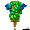
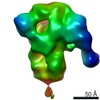
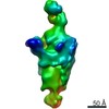
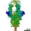
- PDB-6xcm: Structure of the SARS-CoV-2 spike glycoprotein in complex with th... -
+ Open data

- Basic information
| Entry | Database: PDB / ID: 6xcm | |||||||||
|---|---|---|---|---|---|---|---|---|---|---|
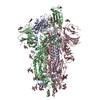
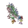
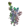
| Title | Structure of the SARS-CoV-2 spike glycoprotein in complex with the C105 neutralizing antibody Fab fragment (state 1) | |||||||||
 Components Components |
| |||||||||
 Keywords Keywords |  VIRAL PROTEIN/IMMUNE SYSTEM / SARS-CoV-2 / Spike glycoprotein / COVID-19 / monoclonal neutralizing antibody / Fabs / nsEMPEM / VIRAL PROTEIN-IMMUNE SYSTEM complex VIRAL PROTEIN/IMMUNE SYSTEM / SARS-CoV-2 / Spike glycoprotein / COVID-19 / monoclonal neutralizing antibody / Fabs / nsEMPEM / VIRAL PROTEIN-IMMUNE SYSTEM complex | |||||||||
| Function / homology |  Function and homology information Function and homology informationMaturation of spike protein / viral translation / Translation of Structural Proteins / Virion Assembly and Release / host cell surface / host extracellular space / suppression by virus of host tetherin activity / Induction of Cell-Cell Fusion / structural constituent of virion / host cell endoplasmic reticulum-Golgi intermediate compartment membrane ...Maturation of spike protein / viral translation / Translation of Structural Proteins / Virion Assembly and Release / host cell surface / host extracellular space / suppression by virus of host tetherin activity / Induction of Cell-Cell Fusion / structural constituent of virion / host cell endoplasmic reticulum-Golgi intermediate compartment membrane / entry receptor-mediated virion attachment to host cell / receptor-mediated endocytosis of virus by host cell / Attachment and Entry / membrane fusion / positive regulation of viral entry into host cell / receptor-mediated virion attachment to host cell / receptor ligand activity / host cell surface receptor binding / fusion of virus membrane with host plasma membrane / fusion of virus membrane with host endosome membrane / viral envelope / symbiont-mediated suppression of host type I interferon-mediated signaling pathway / virion attachment to host cell / SARS-CoV-2 activates/modulates innate and adaptive immune responses / host cell plasma membrane / virion membrane / membrane / identical protein binding / plasma membraneSimilarity search - Function | |||||||||
| Biological species |   Severe acute respiratory syndrome coronavirus 2 Severe acute respiratory syndrome coronavirus 2 Homo sapiens (human) Homo sapiens (human) | |||||||||
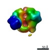
| Method | ELECTRON MICROSCOPY / single particle reconstruction / cryo EM / Resolution: 3.42 Å | |||||||||
 Authors Authors | Barnes, C.O. / Bjorkman, P.J. | |||||||||
| Funding support |  United States, 1items United States, 1items
| |||||||||
 Citation Citation | Journal: Cell / Year: 2020 Title: Structures of Human Antibodies Bound to SARS-CoV-2 Spike Reveal Common Epitopes and Recurrent Features of Antibodies. Authors: Christopher O Barnes / Anthony P West / Kathryn E Huey-Tubman / Magnus A G Hoffmann / Naima G Sharaf / Pauline R Hoffman / Nicholas Koranda / Harry B Gristick / Christian Gaebler / Frauke ...Authors: Christopher O Barnes / Anthony P West / Kathryn E Huey-Tubman / Magnus A G Hoffmann / Naima G Sharaf / Pauline R Hoffman / Nicholas Koranda / Harry B Gristick / Christian Gaebler / Frauke Muecksch / Julio C Cetrulo Lorenzi / Shlomo Finkin / Thomas Hägglöf / Arlene Hurley / Katrina G Millard / Yiska Weisblum / Fabian Schmidt / Theodora Hatziioannou / Paul D Bieniasz / Marina Caskey / Davide F Robbiani / Michel C Nussenzweig / Pamela J Bjorkman / Abstract: Neutralizing antibody responses to coronaviruses mainly target the receptor-binding domain (RBD) of the trimeric spike. Here, we characterized polyclonal immunoglobulin Gs (IgGs) and Fabs from COVID- ...Neutralizing antibody responses to coronaviruses mainly target the receptor-binding domain (RBD) of the trimeric spike. Here, we characterized polyclonal immunoglobulin Gs (IgGs) and Fabs from COVID-19 convalescent individuals for recognition of coronavirus spikes. Plasma IgGs differed in their focus on RBD epitopes, recognition of alpha- and beta-coronaviruses, and contributions of avidity to increased binding/neutralization of IgGs over Fabs. Using electron microscopy, we examined specificities of polyclonal plasma Fabs, revealing recognition of both S1 and RBD epitopes on SARS-CoV-2 spike. Moreover, a 3.4 Å cryo-electron microscopy (cryo-EM) structure of a neutralizing monoclonal Fab-spike complex revealed an epitope that blocks ACE2 receptor binding. Modeling based on these structures suggested different potentials for inter-spike crosslinking by IgGs on viruses, and characterized IgGs would not be affected by identified SARS-CoV-2 spike mutations. Overall, our studies structurally define a recurrent anti-SARS-CoV-2 antibody class derived from VH3-53/VH3-66 and similarity to a SARS-CoV VH3-30 antibody, providing criteria for evaluating vaccine-elicited antibodies. | |||||||||
| History |
|
- Structure visualization
| Movie |
Movie viewer |
|---|---|
| Structure viewer | Molecule: MolmilJmol/JSmol |
- Downloads & links
-Download
| PDBx/mmCIF format | 6xcm.cif.gz | 612.8 KB | Display | PDBx/mmCIF format |
|---|---|---|---|---|
| PDB format | pdb6xcm.ent.gz | 506.1 KB | Display | PDB format |
| PDBx/mmJSON format | 6xcm.json.gz | Tree view | PDBx/mmJSON format | |
| Others |  Other downloads Other downloads |
-Validation report
| Arichive directory | https://data.pdbj.org/pub/pdb/validation_reports/xc/6xcmftp://data.pdbj.org/pub/pdb/validation_reports/xc/6xcm | HTTPS FTP |
|---|
-Related structure data
| Related structure data | 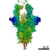 22127MC 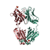 6xcaC 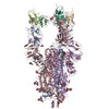 6xcnC M: map data used to model this data C: citing same article ( |
|---|---|
| Similar structure data |


-Links
 PDBj
PDBj


- Assembly
| Deposited unit | 
|
|---|---|
| 1 |
|
-Components
-Protein , 1 types, 3 molecules ABC
| #1: Protein | Spike protein / S glycoprotein / E2 / Peplomer protein Mass: 139788.953 Da / Num. of mol.: 3 Source method: isolated from a genetically manipulated source Source: (gene. exp.) Severe acute respiratory syndrome coronavirus 2Gene: S, 2 / Plasmid: pCAGGS / Cell line (production host): HeLa / Production host: Homo sapiens (human) / Strain (production host): Expi293F / References: UniProt: P0DTC2 |
|---|
-Antibody , 2 types, 4 molecules HNLS
| #2: Antibody | Mass: 24662.506 Da / Num. of mol.: 2 Source method: isolated from a genetically manipulated source Source: (gene. exp.) Homo sapiens (human) / Plasmid: p3BNC / Cell line (production host): HeLa / Production host: Homo sapiens (human) / Strain (production host): Expi293F#3: Antibody | Mass: 22893.227 Da / Num. of mol.: 2 Source method: isolated from a genetically manipulated source Source: (gene. exp.) Homo sapiens (human) / Plasmid: p3BNC / Cell line (production host): HeLa / Production host: Homo sapiens (human) / Strain (production host): Expi293F |
|---|
-Sugars , 5 types, 43 molecules 
| #4: Polysaccharide | 2-acetamido-2-deoxy-beta-D-glucopyranose-(1-4)-2-acetamido-2-deoxy-beta-D-glucopyranose / Mass: 424.401 Da / Num. of mol.: 9Source method: isolated from a genetically manipulated source #5: Polysaccharide | alpha-L-fucopyranose-(1-6)-2-acetamido-2-deoxy-beta-D-glucopyranose | / Mass: 367.349 Da / Num. of mol.: 1Source method: isolated from a genetically manipulated source #6: Polysaccharide | / Mass: 570.542 Da / Num. of mol.: 2Source method: isolated from a genetically manipulated source #7: Polysaccharide | beta-D-mannopyranose-(1-4)-2-acetamido-2-deoxy-beta-D-glucopyranose-(1-4)-2-acetamido-2-deoxy-beta- ...beta-D-mannopyranose-(1-4)-2-acetamido-2-deoxy-beta-D-glucopyranose-(1-4)-2-acetamido-2-deoxy-beta-D-glucopyranose | / Mass: 586.542 Da / Num. of mol.: 1Source method: isolated from a genetically manipulated source #8: Sugar | ChemComp-NAG / N-Acetylglucosamine Type: D-saccharide, beta linking / Mass: 221.208 Da / Num. of mol.: 30 Type: D-saccharide, beta linking / Mass: 221.208 Da / Num. of mol.: 30Source method: isolated from a genetically manipulated source Formula: C8H15NO6 |
|---|
-Details
| Has ligand of interest | N |
|---|
-Experimental details
-Experiment
| Experiment | Method: ELECTRON MICROSCOPY |
|---|---|
| EM experiment | Aggregation state: PARTICLE / 3D reconstruction method: single particle reconstruction |
- Sample preparation
| Component |
| ||||||||||||||||||||||||
|---|---|---|---|---|---|---|---|---|---|---|---|---|---|---|---|---|---|---|---|---|---|---|---|---|---|
| Molecular weight | Value: 0.7 MDa / Experimental value: YES | ||||||||||||||||||||||||
| Source (natural) |
| ||||||||||||||||||||||||
| Source (recombinant) |
| ||||||||||||||||||||||||
| Buffer solution | pH: 8 | ||||||||||||||||||||||||
| Buffer component |
| ||||||||||||||||||||||||
| Specimen | Conc.: 1.5 mg/ml / Embedding applied: NO / Shadowing applied: NO / Staining applied: NO / Vitrification applied: YES Details: Monodisperse sample taken after size-exclusion chromatography | ||||||||||||||||||||||||
| Specimen support | Details: 60 s glow discharge in air at 0.20 mA current / Grid material: GOLD / Grid mesh size: 300 divisions/in. / Grid type: UltrAuFoil R1.2/1.3 | ||||||||||||||||||||||||
| Vitrification | Instrument: FEI VITROBOT MARK IV / Cryogen name: ETHANE / Humidity: 100 % / Chamber temperature: 295 K Details: 3 microliters added, blotted for 3 s with 0 blot force |
- Electron microscopy imaging
| Experimental equipment |  Model: Titan Krios / Image courtesy: FEI Company |
|---|---|
| Microscopy | Model: FEI TITAN KRIOS / Details: Collected with 3x3 pattern and beam image shift |
| Electron gun | Electron source: FIELD EMISSION GUN / Accelerating voltage: 300 kV / Illumination mode: FLOOD BEAM |
| Electron lens | Mode: BRIGHT FIELDBright-field microscopy / Nominal magnification: 105000 X / Nominal defocus max: 2500 nm / Nominal defocus min: 1000 nm / Cs: 2.7 mm / Alignment procedure: COMA FREE |
| Specimen holder | Cryogen: NITROGEN / Specimen holder model: FEI TITAN KRIOS AUTOGRID HOLDER |
| Image recording | Average exposure time: 1.89 sec. / Electron dose: 60 e/Å2 / Film or detector model: GATAN K3 BIOQUANTUM (6k x 4k) / Num. of grids imaged: 1 / Num. of real images: 5940 |
- Processing
| Software | Name: PHENIX / Version: 1.17.1_3660: / Classification: refinement | ||||||||||||||||||||||||||||||||||||
|---|---|---|---|---|---|---|---|---|---|---|---|---|---|---|---|---|---|---|---|---|---|---|---|---|---|---|---|---|---|---|---|---|---|---|---|---|---|
| EM software |
| ||||||||||||||||||||||||||||||||||||
| Image processing | Details: Gain-normalized and collected in counting mode with 40 frames per movie | ||||||||||||||||||||||||||||||||||||
| CTF correction | Type: PHASE FLIPPING AND AMPLITUDE CORRECTION | ||||||||||||||||||||||||||||||||||||
| Symmetry | Point symmetry: C1 (asymmetric) | ||||||||||||||||||||||||||||||||||||
| 3D reconstruction | Resolution: 3.42 Å / Resolution method: FSC 0.143 CUT-OFF / Num. of particles: 58873 / Symmetry type: POINT | ||||||||||||||||||||||||||||||||||||
| Atomic model building | B value: 90 / Protocol: RIGID BODY FIT / Space: REAL / Target criteria: Correlation coefficient | ||||||||||||||||||||||||||||||||||||
| Atomic model building | PDB-ID: 6VYB Pdb chain-ID: B | ||||||||||||||||||||||||||||||||||||
| Refine LS restraints |
|