 Movie
Movie Controller

[English] 日本語
 Yorodumi
- PDB-6ue0: Crystal structure of dihydrodipicolinate synthase from Klebsiella... -
+ Open data

- Basic information
| Entry | Database: PDB / ID: 6ue0 | ||||||||||||
|---|---|---|---|---|---|---|---|---|---|---|---|---|---|
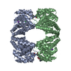
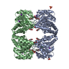
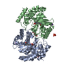
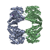
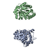
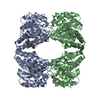
| Title | Crystal structure of dihydrodipicolinate synthase from Klebsiella pneumoniae bound to pyruvate | ||||||||||||
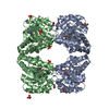
 Components Components | 4-hydroxy-tetrahydrodipicolinate synthase Dihydrodipicolinate synthase Dihydrodipicolinate synthase | ||||||||||||
 Keywords Keywords | LYASE / DHDPS / Lysine biosynthesis / tetramer | ||||||||||||
| Function / homology |  Function and homology information4-hydroxy-tetrahydrodipicolinate synthase / 4-hydroxy-tetrahydrodipicolinate synthase activity / diaminopimelate biosynthetic process / lysine biosynthetic process via diaminopimelate / cytoplasm Function and homology information4-hydroxy-tetrahydrodipicolinate synthase / 4-hydroxy-tetrahydrodipicolinate synthase activity / diaminopimelate biosynthetic process / lysine biosynthetic process via diaminopimelate / cytoplasmSimilarity search - Function | ||||||||||||
| Biological species |  Klebsiella pneumoniae (bacteria) Klebsiella pneumoniae (bacteria) | ||||||||||||
| Method | X-RAY DIFFRACTION / SYNCHROTRON / MOLECULAR REPLACEMENT / Resolution: 1.892 Å | ||||||||||||
 Authors Authors | Impey, R.E. / Lee, M. / Hawkins, D.A. / Sutton, J.M. / Panjikar, S. / Perugini, M.A. / Soares da Costa, T.P. | ||||||||||||
| Funding support |  Australia, 3items Australia, 3items
| ||||||||||||
 Citation Citation | Journal: Febs Lett. / Year: 2020 Title: Mis-annotations of a promising antibiotic target in high-priority gram-negative pathogens. Authors: Impey, R.E. / Lee, M. / Hawkins, D.A. / Sutton, J.M. / Panjikar, S. / Perugini, M.A. / Soares da Costa, T.P. | ||||||||||||
| History |
|
- Structure visualization
| Structure viewer | Molecule: MolmilJmol/JSmol |
|---|
- Downloads & links
-Download
| PDBx/mmCIF format | 6ue0.cif.gz | 138 KB | Display | PDBx/mmCIF format |
|---|---|---|---|---|
| PDB format | pdb6ue0.ent.gz | Display | PDB format | |
| PDBx/mmJSON format | 6ue0.json.gz | Tree view | PDBx/mmJSON format | |
| Others |  Other downloads Other downloads |
-Validation report
| Arichive directory | https://data.pdbj.org/pub/pdb/validation_reports/ue/6ue0ftp://data.pdbj.org/pub/pdb/validation_reports/ue/6ue0 | HTTPS FTP |
|---|
-Related structure data
| Related structure data |  1yxcS S: Starting model for refinement |
|---|---|
| Similar structure data |


-Links
 PDBj
PDBj- Assembly
| Deposited unit | 
| ||||||||
|---|---|---|---|---|---|---|---|---|---|
| 1 | 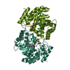
| ||||||||
| Unit cell |
|
-Components
| #1: Protein | Dihydrodipicolinate synthase / HTPA synthase Mass: 34816.828 Da / Num. of mol.: 2 Source method: isolated from a genetically manipulated source Source: (gene. exp.) Klebsiella pneumoniae (bacteria) / Gene: dapA_2, dapA_3, dapA_4, dapA_5, dapA_6, dapA_7 / Production host: Escherichia coli BL21(DE3) (bacteria) / Strain (production host): BL21(DE3)References: UniProt: W9BBZ5, 4-hydroxy-tetrahydrodipicolinate synthase#2: Chemical | Sulfate  Mass: 96.063 Da / Num. of mol.: 2 / Source method: obtained synthetically / Formula: SO4 Mass: 96.063 Da / Num. of mol.: 2 / Source method: obtained synthetically / Formula: SO4#3: Chemical | ChemComp-CL / Chloride  Mass: 35.453 Da / Num. of mol.: 6 / Source method: obtained synthetically / Formula: Cl Mass: 35.453 Da / Num. of mol.: 6 / Source method: obtained synthetically / Formula: Cl#4: Water | ChemComp-HOH / | Water Mass: 18.015 Da / Num. of mol.: 423 / Source method: isolated from a natural source / Formula: H2O Mass: 18.015 Da / Num. of mol.: 423 / Source method: isolated from a natural source / Formula: H2OHas ligand of interest | N | |
|---|
-Experimental details
-Experiment
| Experiment | Method: X-RAY DIFFRACTION / Number of used crystals: 1 |
|---|
- Sample preparation
| Crystal | Density Matthews: 2.4 Å3/Da / Density % sol: 48.66 % |
|---|---|
| Crystal grow | Temperature: 293 K / Method: vapor diffusion, hanging drop Details: 0.1 M Tris, ph 7.5, 2 M Ammonium Sulfate, 5 mM Pyruvate |
-Data collection
| Diffraction | Mean temperature: 100 K / Serial crystal experiment: N |
|---|---|
| Diffraction source | Source: SYNCHROTRON / Site: Australian Synchrotron / Beamline: MX2 / Wavelength: 0.9537 Å |
| Detector | Type: DECTRIS EIGER X 16M / Detector: PIXEL / Date: Mar 8, 2018 |
| Radiation | Protocol: SINGLE WAVELENGTH / Monochromatic (M) / Laue (L): M / Scattering type: x-ray |
| Radiation wavelength | Wavelength: 0.9537 Å / Relative weight: 1 |
| Reflection | Resolution: 1.89→49.039 Å / Num. obs: 54112 / % possible obs: 99.7 % / Redundancy: 6.7 % / CC1/2: 0.998 / Net I/σ(I): 11.4 |
| Reflection shell | Resolution: 1.89→1.93 Å / Num. unique obs: 3259 / CC1/2: 0.827 / % possible all: 95.3 |
- Processing
| Software |
| ||||||||||||||||||||||||||||||||||||||||||||||||||||||||||||||||||||||||||||||||||||||||||||||||||||||||||||||||||||||||||||||||||||||||||||||||||||||
|---|---|---|---|---|---|---|---|---|---|---|---|---|---|---|---|---|---|---|---|---|---|---|---|---|---|---|---|---|---|---|---|---|---|---|---|---|---|---|---|---|---|---|---|---|---|---|---|---|---|---|---|---|---|---|---|---|---|---|---|---|---|---|---|---|---|---|---|---|---|---|---|---|---|---|---|---|---|---|---|---|---|---|---|---|---|---|---|---|---|---|---|---|---|---|---|---|---|---|---|---|---|---|---|---|---|---|---|---|---|---|---|---|---|---|---|---|---|---|---|---|---|---|---|---|---|---|---|---|---|---|---|---|---|---|---|---|---|---|---|---|---|---|---|---|---|---|---|---|---|---|---|
| Refinement | Method to determine structure: MOLECULAR REPLACEMENT Starting model: 1YXC Resolution: 1.892→48.99 Å / Cor.coef. Fo:Fc: 0.96 / Cor.coef. Fo:Fc free: 0.947 / SU B: 2.582 / SU ML: 0.076 / Cross valid method: FREE R-VALUE / ESU R: 0.126 / ESU R Free: 0.117 Details: Hydrogens have been added in their riding positions
| ||||||||||||||||||||||||||||||||||||||||||||||||||||||||||||||||||||||||||||||||||||||||||||||||||||||||||||||||||||||||||||||||||||||||||||||||||||||
| Solvent computation | Ion probe radii: 0.8 Å / Shrinkage radii: 0.8 Å / VDW probe radii: 1.2 Å | ||||||||||||||||||||||||||||||||||||||||||||||||||||||||||||||||||||||||||||||||||||||||||||||||||||||||||||||||||||||||||||||||||||||||||||||||||||||
| Displacement parameters | Biso mean: 22.372 Å2
| ||||||||||||||||||||||||||||||||||||||||||||||||||||||||||||||||||||||||||||||||||||||||||||||||||||||||||||||||||||||||||||||||||||||||||||||||||||||
| Refinement step | Cycle: LAST / Resolution: 1.892→48.99 Å
| ||||||||||||||||||||||||||||||||||||||||||||||||||||||||||||||||||||||||||||||||||||||||||||||||||||||||||||||||||||||||||||||||||||||||||||||||||||||
| Refine LS restraints |
| ||||||||||||||||||||||||||||||||||||||||||||||||||||||||||||||||||||||||||||||||||||||||||||||||||||||||||||||||||||||||||||||||||||||||||||||||||||||
| LS refinement shell |
|
