 Movie
Movie Controller


+ Open data

- Basic information
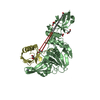
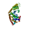
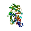
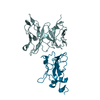
| Entry | Database: PDB / ID: 6tur | ||||||
|---|---|---|---|---|---|---|---|
| Title | human XPG, Apo1 form | ||||||
 Components Components | DNA repair protein complementing XP-G cells,DNA repair protein complementing XP-G cells | ||||||
 Keywords Keywords | DNA BINDING PROTEIN / XPG nuclease domain | ||||||
| Function / homology |  Function and homology information Function and homology informationnucleotide-excision repair complex / base-excision repair, AP site formation / bubble DNA binding / regulation of catalytic activity / response to UV-C / RNA polymerase II complex binding / enzyme activator activity / transcription-coupled nucleotide-excision repair / response to UV / DNA endonuclease activity ...nucleotide-excision repair complex / base-excision repair, AP site formation / bubble DNA binding / regulation of catalytic activity / response to UV-C / RNA polymerase II complex binding / enzyme activator activity / transcription-coupled nucleotide-excision repair / response to UV / DNA endonuclease activity / nucleotide-excision repair / double-strand break repair via homologous recombination / Dual Incision in GG-NER / Formation of Incision Complex in GG-NER / Dual incision in TC-NER / single-stranded DNA binding / chromosome / double-stranded DNA binding / endonuclease activity / damaged DNA binding / Hydrolases; Acting on ester bonds / protein-containing complex binding / negative regulation of apoptotic process / protein homodimerization activity / protein-containing complex / nucleoplasm / metal ion binding / nucleusSimilarity search - Function | ||||||
| Biological species |  Homo sapiens (human) Homo sapiens (human) | ||||||
| Method | X-RAY DIFFRACTION / SYNCHROTRON / SAD / Resolution: 2.9 Å | ||||||
 Authors Authors | Ruiz, F.M. / Fernandez-Tornero, C. | ||||||
 Citation Citation | Journal: Nucleic Acids Res. / Year: 2020 Title: The crystal structure of human XPG, the xeroderma pigmentosum group G endonuclease, provides insight into nucleotide excision DNA repair. Authors: Gonzalez-Corrochano, R. / Ruiz, F.M. / Taylor, N.M.I. / Huecas, S. / Drakulic, S. / Spinola-Amilibia, M. / Fernandez-Tornero, C. | ||||||
| History |
|
- Structure visualization
| Structure viewer | Molecule: MolmilJmol/JSmol |
|---|
- Downloads & links
-Download
| PDBx/mmCIF format | 6tur.cif.gz | 268.8 KB | Display | PDBx/mmCIF format |
|---|---|---|---|---|
| PDB format | pdb6tur.ent.gz | Display | PDB format | |
| PDBx/mmJSON format | 6tur.json.gz | Tree view | PDBx/mmJSON format | |
| Others |  Other downloads Other downloads |
-Validation report
| Arichive directory | https://data.pdbj.org/pub/pdb/validation_reports/tu/6turftp://data.pdbj.org/pub/pdb/validation_reports/tu/6tur | HTTPS FTP |
|---|
-Related structure data


-Links
 PDBj
PDBj

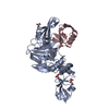
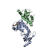
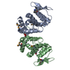
- Assembly
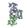
| Deposited unit | 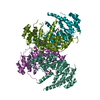
| ||||||||||||||||||||||||||||
|---|---|---|---|---|---|---|---|---|---|---|---|---|---|---|---|---|---|---|---|---|---|---|---|---|---|---|---|---|---|
| 1 | 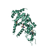
| ||||||||||||||||||||||||||||
| 2 | 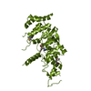
| ||||||||||||||||||||||||||||
| 3 | 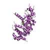
| ||||||||||||||||||||||||||||
| 4 | 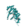
| ||||||||||||||||||||||||||||
| Unit cell |
| ||||||||||||||||||||||||||||
| Noncrystallographic symmetry (NCS) | NCS domain:
NCS ensembles :
|
-Components
| #1: Protein | / DNA excision repair protein ERCC-5 / Xeroderma pigmentosum group G-complementing protein Mass: 40977.465 Da / Num. of mol.: 4 Source method: isolated from a genetically manipulated source Source: (gene. exp.) Homo sapiens (human) / Gene: ERCC5, ERCM2, XPG, XPGC / Production host:  Escherichia coli (E. coli) Escherichia coli (E. coli)References: UniProt: P28715, Hydrolases; Acting on ester bonds |
|---|
-Experimental details
-Experiment
| Experiment | Method: X-RAY DIFFRACTION / Number of used crystals: 1 |
|---|
- Sample preparation
| Crystal | Density Matthews: 2.87 Å3/Da / Density % sol: 57.21 % |
|---|---|
| Crystal grow | Temperature: 295 K / Method: vapor diffusion, sitting drop / Details: 25% PEG 3350, 100 mM Citric acid pH 3.5 |
-Data collection
| Diffraction | Mean temperature: 100 K / Serial crystal experiment: N |
|---|---|
| Diffraction source | Source: SYNCHROTRON / Site: ALBA  / Beamline: XALOC / Wavelength: 0.9786 Å / Beamline: XALOC / Wavelength: 0.9786 Å |
| Detector | Type: DECTRIS PILATUS 6M / Detector: PIXEL / Date: Feb 13, 2013 |
| Radiation | Protocol: SINGLE WAVELENGTH / Monochromatic (M) / Laue (L): M / Scattering type: x-ray |
| Radiation wavelength | Wavelength: 0.9786 Å / Relative weight: 1 |
| Reflection | Resolution: 2.9→134.475 Å / Num. obs: 42886 / % possible obs: 100 % / Redundancy: 80 % / CC1/2: 0.998 / Net I/σ(I): 13.1 |
| Reflection shell | Resolution: 2.9→3.01 Å / Num. unique obs: 4403 / CC1/2: 0.528 |
- Processing
| Software |
| ||||||||||||||||||||||||||||||||||||||||||||||||||||||||||||||||||||||||||||||||||||||||||||||||||||||||||||||||||||||||||||||||||||||||||||||||||||||||||||||||||||||||||||||||||||||
|---|---|---|---|---|---|---|---|---|---|---|---|---|---|---|---|---|---|---|---|---|---|---|---|---|---|---|---|---|---|---|---|---|---|---|---|---|---|---|---|---|---|---|---|---|---|---|---|---|---|---|---|---|---|---|---|---|---|---|---|---|---|---|---|---|---|---|---|---|---|---|---|---|---|---|---|---|---|---|---|---|---|---|---|---|---|---|---|---|---|---|---|---|---|---|---|---|---|---|---|---|---|---|---|---|---|---|---|---|---|---|---|---|---|---|---|---|---|---|---|---|---|---|---|---|---|---|---|---|---|---|---|---|---|---|---|---|---|---|---|---|---|---|---|---|---|---|---|---|---|---|---|---|---|---|---|---|---|---|---|---|---|---|---|---|---|---|---|---|---|---|---|---|---|---|---|---|---|---|---|---|---|---|---|
| Refinement | Method to determine structure: SAD / Resolution: 2.9→67.6 Å / Cor.coef. Fo:Fc: 0.923 / Cor.coef. Fo:Fc free: 0.894 / SU B: 25.688 / SU ML: 0.433 / Cross valid method: FREE R-VALUE / ESU R: 2.713 / ESU R Free: 0.408 Details: Hydrogens have been added in their riding positions
| ||||||||||||||||||||||||||||||||||||||||||||||||||||||||||||||||||||||||||||||||||||||||||||||||||||||||||||||||||||||||||||||||||||||||||||||||||||||||||||||||||||||||||||||||||||||
| Solvent computation | Ion probe radii: 0.8 Å / Shrinkage radii: 0.8 Å / VDW probe radii: 1.2 Å / Solvent model: MASK BULK SOLVENT | ||||||||||||||||||||||||||||||||||||||||||||||||||||||||||||||||||||||||||||||||||||||||||||||||||||||||||||||||||||||||||||||||||||||||||||||||||||||||||||||||||||||||||||||||||||||
| Displacement parameters | Biso mean: 101.744 Å2
| ||||||||||||||||||||||||||||||||||||||||||||||||||||||||||||||||||||||||||||||||||||||||||||||||||||||||||||||||||||||||||||||||||||||||||||||||||||||||||||||||||||||||||||||||||||||
| Refinement step | Cycle: LAST / Resolution: 2.9→67.6 Å
| ||||||||||||||||||||||||||||||||||||||||||||||||||||||||||||||||||||||||||||||||||||||||||||||||||||||||||||||||||||||||||||||||||||||||||||||||||||||||||||||||||||||||||||||||||||||
| Refine LS restraints |
|