 Movie
Movie Controller

[English] 日本語
 Yorodumi
- PDB-6tef: Crystal structure of monooxygenase RutA complexed with dioxygen u... -
+ Open data

- Basic information
| Entry | Database: PDB / ID: 6tef | |||||||||
|---|---|---|---|---|---|---|---|---|---|---|
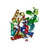
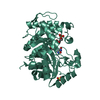
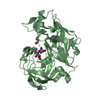
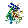
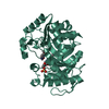
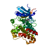
| Title | Crystal structure of monooxygenase RutA complexed with dioxygen under 0.5 MPa / 5 bars of oxygen pressure. | |||||||||
 Components Components | Pyrimidine monooxygenase RutA | |||||||||
 Keywords Keywords |  FLAVOPROTEIN / monooxygenase / RutA / FMN / flavin-N5-oxide / bioengineering FLAVOPROTEIN / monooxygenase / RutA / FMN / flavin-N5-oxide / bioengineering | |||||||||
| Function / homology |  Function and homology informationpyrimidine oxygenase / uracil oxygenase activity / pyrimidine nucleobase catabolic process / alkanesulfonate monooxygenase activity / alkanesulfonate catabolic process / thymine catabolic process / uracil catabolic process / nitrogen utilization / monooxygenase activity Function and homology informationpyrimidine oxygenase / uracil oxygenase activity / pyrimidine nucleobase catabolic process / alkanesulfonate monooxygenase activity / alkanesulfonate catabolic process / thymine catabolic process / uracil catabolic process / nitrogen utilization / monooxygenase activitySimilarity search - Function | |||||||||
| Biological species |  Escherichia coli K-12 (bacteria) Escherichia coli K-12 (bacteria) | |||||||||
| Method | X-RAY DIFFRACTION / SYNCHROTRON / MOLECULAR REPLACEMENT / Resolution: 1.8 Å | |||||||||
 Authors Authors | Saleem-Batcha, R. / Matthews, A. / Teufel, R. | |||||||||
| Funding support |  Germany, 2items Germany, 2items
| |||||||||
 Citation Citation | Journal: Nat.Chem.Biol. / Year: 2020 Title: Aminoperoxide adducts expand the catalytic repertoire of flavin monooxygenases. Authors: Matthews, A. / Saleem-Batcha, R. / Sanders, J.N. / Stull, F. / Houk, K.N. / Teufel, R. | |||||||||
| History |
|
- Structure visualization
| Structure viewer | Molecule: MolmilJmol/JSmol |
|---|
- Downloads & links
-Download
| PDBx/mmCIF format | 6tef.cif.gz | 90.5 KB | Display | PDBx/mmCIF format |
|---|---|---|---|---|
| PDB format | pdb6tef.ent.gz | Display | PDB format | |
| PDBx/mmJSON format | 6tef.json.gz | Tree view | PDBx/mmJSON format | |
| Others |  Other downloads Other downloads |
-Validation report
| Arichive directory | https://data.pdbj.org/pub/pdb/validation_reports/te/6tefftp://data.pdbj.org/pub/pdb/validation_reports/te/6tef | HTTPS FTP |
|---|
-Related structure data
| Related structure data | 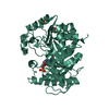 6sggC  6sglC 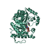 6sgmC  6sgnC 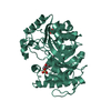 6teeC  6tegC  5wanS C: citing same article ( S: Starting model for refinement |
|---|---|
| Similar structure data |


-Links
 PDBj
PDBj


- Assembly
| Deposited unit | 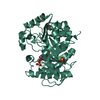
| ||||||||
|---|---|---|---|---|---|---|---|---|---|
| 1 |
| ||||||||
| Unit cell |
|
-Components
-Protein , 1 types, 1 molecules AAA
| #1: Protein | Mass: 40111.336 Da / Num. of mol.: 1 Source method: isolated from a genetically manipulated source Source: (gene. exp.) Escherichia coli K-12 (bacteria) / Gene: rutA, ycdM, b1012, JW0997 / Production host: Escherichia coli BL21(DE3) (bacteria) / References: UniProt: P75898, pyrimidine oxygenase |
|---|
-Non-polymers , 5 types, 207 molecules 


| #2: Chemical | Sulfate Mass: 96.063 Da / Num. of mol.: 3 / Source method: obtained synthetically / Formula: SO4 Mass: 96.063 Da / Num. of mol.: 3 / Source method: obtained synthetically / Formula: SO4#3: Chemical | ChemComp-GOL / | Glycerol Mass: 92.094 Da / Num. of mol.: 1 / Source method: obtained synthetically / Formula: C3H8O3 Mass: 92.094 Da / Num. of mol.: 1 / Source method: obtained synthetically / Formula: C3H8O3#4: Chemical | ChemComp-FMN / | Flavin mononucleotide Mass: 456.344 Da / Num. of mol.: 1 / Source method: obtained synthetically / Formula: C17H21N4O9P / Feature type: SUBJECT OF INVESTIGATION Mass: 456.344 Da / Num. of mol.: 1 / Source method: obtained synthetically / Formula: C17H21N4O9P / Feature type: SUBJECT OF INVESTIGATION#5: Chemical | ChemComp-OXY / | Oxygen Mass: 31.999 Da / Num. of mol.: 1 / Source method: obtained synthetically / Formula: O2 Mass: 31.999 Da / Num. of mol.: 1 / Source method: obtained synthetically / Formula: O2#6: Water | ChemComp-HOH / | WaterMass: 18.015 Da / Num. of mol.: 201 / Source method: isolated from a natural source / Formula: H2O |
|---|
-Details
| Has ligand of interest | Y |
|---|
-Experimental details
-Experiment
| Experiment | Method: X-RAY DIFFRACTION / Number of used crystals: 1 |
|---|
- Sample preparation
| Crystal | Density Matthews: 2.67 Å3/Da / Density % sol: 53.95 % |
|---|---|
| Crystal grow | Temperature: 295 K / Method: vapor diffusion, sitting drop Details: 100 mM Bis-Tris pH 6.5, 1900 mM Ammonium Sulphate, 2-5% MPD (v/v), 1 mM FMN |
-Data collection
| Diffraction | Mean temperature: 100 K / Serial crystal experiment: N |
|---|---|
| Diffraction source | Source: SYNCHROTRON / Site: SLS  / Beamline: X06SA / Wavelength: 1 Å / Beamline: X06SA / Wavelength: 1 Å |
| Detector | Type: DECTRIS EIGER X 16M / Detector: PIXEL / Date: Jun 27, 2018 |
| Radiation | Protocol: SINGLE WAVELENGTH / Monochromatic (M) / Laue (L): M / Scattering type: x-ray |
| Radiation wavelength | Wavelength: 1 Å / Relative weight: 1 |
| Reflection | Resolution: 1.8→48.29 Å / Num. obs: 40277 / % possible obs: 100 % / Redundancy: 9.9 % / Rmerge(I) obs: 0.08 / Net I/σ(I): 17.2 |
| Reflection shell | Resolution: 1.8→1.9 Å / Num. unique obs: 5809 / CC1/2: 0.84 |
- Processing
| Software |
| ||||||||||||||||||||||||||||||||||||||||||||||||||||||||||||||||||||||||||||||||||||||||||||||||||||||||||||||||||||||||||||||||||||||||||||||||||||||
|---|---|---|---|---|---|---|---|---|---|---|---|---|---|---|---|---|---|---|---|---|---|---|---|---|---|---|---|---|---|---|---|---|---|---|---|---|---|---|---|---|---|---|---|---|---|---|---|---|---|---|---|---|---|---|---|---|---|---|---|---|---|---|---|---|---|---|---|---|---|---|---|---|---|---|---|---|---|---|---|---|---|---|---|---|---|---|---|---|---|---|---|---|---|---|---|---|---|---|---|---|---|---|---|---|---|---|---|---|---|---|---|---|---|---|---|---|---|---|---|---|---|---|---|---|---|---|---|---|---|---|---|---|---|---|---|---|---|---|---|---|---|---|---|---|---|---|---|---|---|---|---|
| Refinement | Method to determine structure: MOLECULAR REPLACEMENT Starting model: 5WAN Resolution: 1.8→43.875 Å / Cor.coef. Fo:Fc: 0.97 / Cor.coef. Fo:Fc free: 0.956 / SU B: 2.032 / SU ML: 0.063 / Cross valid method: THROUGHOUT / ESU R: 0.097 / ESU R Free: 0.099 Details: Hydrogens have been added in their riding positions
| ||||||||||||||||||||||||||||||||||||||||||||||||||||||||||||||||||||||||||||||||||||||||||||||||||||||||||||||||||||||||||||||||||||||||||||||||||||||
| Solvent computation | Ion probe radii: 0.8 Å / Shrinkage radii: 0.8 Å / VDW probe radii: 1.2 Å | ||||||||||||||||||||||||||||||||||||||||||||||||||||||||||||||||||||||||||||||||||||||||||||||||||||||||||||||||||||||||||||||||||||||||||||||||||||||
| Displacement parameters | Biso mean: 27.675 Å2
| ||||||||||||||||||||||||||||||||||||||||||||||||||||||||||||||||||||||||||||||||||||||||||||||||||||||||||||||||||||||||||||||||||||||||||||||||||||||
| Refinement step | Cycle: LAST / Resolution: 1.8→43.875 Å
| ||||||||||||||||||||||||||||||||||||||||||||||||||||||||||||||||||||||||||||||||||||||||||||||||||||||||||||||||||||||||||||||||||||||||||||||||||||||
| Refine LS restraints |
| ||||||||||||||||||||||||||||||||||||||||||||||||||||||||||||||||||||||||||||||||||||||||||||||||||||||||||||||||||||||||||||||||||||||||||||||||||||||
| LS refinement shell |
|
