 Movie
Movie Controller


+ Open data

- Basic information
| Entry | Database: PDB / ID: 6t9r | ||||||
|---|---|---|---|---|---|---|---|
| Title | Aplysia californica AChBP in complex with a cytisine derivative | ||||||
 Components Components | Acetylcholine binding protein | ||||||
 Keywords Keywords |  HYDROLASE / Acetylcholine binding protein / nicotinic receptor surrogate HYDROLASE / Acetylcholine binding protein / nicotinic receptor surrogate | ||||||
| Function / homology |  Function and homology information Function and homology informationextracellular ligand-gated monoatomic ion channel activity / transmembrane signaling receptor activity / membrane / identical protein binding / metal ion bindingSimilarity search - Function | ||||||
| Biological species |  Aplysia californica (California sea hare) Aplysia californica (California sea hare) | ||||||
| Method | X-RAY DIFFRACTION / SYNCHROTRON / MOLECULAR REPLACEMENT / Resolution: 1.72 Å | ||||||
 Authors Authors | Davis, S. / Hunter, W.N. | ||||||
| Funding support |  United Kingdom, 1items United Kingdom, 1items
| ||||||
 Citation Citation | Journal: Acta Crystallogr.,Sect.F / Year: 2020 Title: The thermodynamic profile and molecular interactions of a C(9)-cytisine derivative-binding acetylcholine-binding protein from Aplysia californica. Authors: Davis, S. / Rego Campello, H. / Gallagher, T. / Hunter, W.N. | ||||||
| History |
|
- Structure visualization
| Structure viewer | Molecule: MolmilJmol/JSmol |
|---|
- Downloads & links
-Download
| PDBx/mmCIF format | 6t9r.cif.gz | 879.9 KB | Display | PDBx/mmCIF format |
|---|---|---|---|---|
| PDB format | pdb6t9r.ent.gz | Display | PDB format | |
| PDBx/mmJSON format | 6t9r.json.gz | Tree view | PDBx/mmJSON format | |
| Others |  Other downloads Other downloads |
-Validation report
| Arichive directory | https://data.pdbj.org/pub/pdb/validation_reports/t9/6t9rftp://data.pdbj.org/pub/pdb/validation_reports/t9/6t9r | HTTPS FTP |
|---|
-Related structure data
| Related structure data | 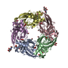 6qkkS S: Starting model for refinement |
|---|---|
| Similar structure data |

-Links
 PDBj
PDBj

- Assembly
| Deposited unit | 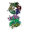
| ||||||||
|---|---|---|---|---|---|---|---|---|---|
| 1 | 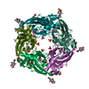
| ||||||||
| 2 | 
| ||||||||
| Unit cell |
| ||||||||
| Components on special symmetry positions |
|
-Components
-Protein / Sugars , 2 types, 20 molecules AAABBBCCCDDDEEEFFFGGGHHHIIIJJJ
| #1: Protein | Mass: 28326.750 Da / Num. of mol.: 10 Source method: isolated from a genetically manipulated source Source: (gene. exp.) Aplysia californica (California sea hare)Plasmid: pFastBac 1 / Production host:  Trichoplusia ni (cabbage looper) / References: UniProt: Q8WSF8 Trichoplusia ni (cabbage looper) / References: UniProt: Q8WSF8#3: Sugar | ChemComp-NAG / N-Acetylglucosamine Type: D-saccharide, beta linking / Mass: 221.208 Da / Num. of mol.: 10 Type: D-saccharide, beta linking / Mass: 221.208 Da / Num. of mol.: 10Source method: isolated from a genetically manipulated source Formula: C8H15NO6 |
|---|
-Non-polymers , 6 types, 2980 molecules 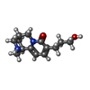


| #2: Chemical | ChemComp-MXQ / (  Mass: 248.321 Da / Num. of mol.: 10 / Source method: obtained synthetically / Formula: C14H20N2O2 / Feature type: SUBJECT OF INVESTIGATION Mass: 248.321 Da / Num. of mol.: 10 / Source method: obtained synthetically / Formula: C14H20N2O2 / Feature type: SUBJECT OF INVESTIGATION#4: Chemical | ChemComp-GOL / Glycerol Mass: 92.094 Da / Num. of mol.: 41 / Source method: obtained synthetically / Formula: C3H8O3 Mass: 92.094 Da / Num. of mol.: 41 / Source method: obtained synthetically / Formula: C3H8O3#5: Chemical | ChemComp-PO4 / Phosphate Mass: 94.971 Da / Num. of mol.: 10 / Source method: obtained synthetically / Formula: PO4 Mass: 94.971 Da / Num. of mol.: 10 / Source method: obtained synthetically / Formula: PO4#6: Chemical | ChemComp-CL / Chloride Mass: 35.453 Da / Num. of mol.: 10 / Source method: obtained synthetically / Formula: Cl Mass: 35.453 Da / Num. of mol.: 10 / Source method: obtained synthetically / Formula: Cl#7: Chemical | ChemComp-K /  Mass: 39.098 Da / Num. of mol.: 10 / Source method: obtained synthetically / Formula: K Mass: 39.098 Da / Num. of mol.: 10 / Source method: obtained synthetically / Formula: K#8: Water | ChemComp-HOH / | WaterMass: 18.015 Da / Num. of mol.: 2899 / Source method: isolated from a natural source / Formula: H2O |
|---|
-Details
| Has ligand of interest | Y |
|---|
-Experimental details
-Experiment
| Experiment | Method: X-RAY DIFFRACTION / Number of used crystals: 1 |
|---|
- Sample preparation
| Crystal | Density Matthews: 3.14 Å3/Da / Density % sol: 60.87 % / Description: Block |
|---|---|
| Crystal grow | Temperature: 295.15 K / Method: vapor diffusion, hanging drop / pH: 7 Details: Starting protein concentration 12.5 mg/ml + 6 mM ligand BS82. Crystallised as hanging drops, the drop containing 1.5 ul protein, 0.2 ul reservoir (0.8 M NaH2PO4, 0.8 M KH2PO4, 10% glycerol, ...Details: Starting protein concentration 12.5 mg/ml + 6 mM ligand BS82. Crystallised as hanging drops, the drop containing 1.5 ul protein, 0.2 ul reservoir (0.8 M NaH2PO4, 0.8 M KH2PO4, 10% glycerol, 0.1 M HEPES pH 7.0) and 0.3 ul microseeds from cytisine bound AChBP crystals (grown as sitting drops in 0.8 M NaH2PO4, 0.8 M KH2PO4, 0.1 M HEPES pH 7.5). |
-Data collection
| Diffraction | Mean temperature: 100 K / Serial crystal experiment: N |
|---|---|
| Diffraction source | Source: SYNCHROTRON / Site: Diamond / Beamline: I03 / Wavelength: 0.976 Å |
| Detector | Type: DECTRIS EIGER2 X 16M / Detector: PIXEL / Date: Aug 4, 2019 |
| Radiation | Protocol: SINGLE WAVELENGTH / Monochromatic (M) / Laue (L): M / Scattering type: x-ray |
| Radiation wavelength | Wavelength: 0.976 Å / Relative weight: 1 |
| Reflection | Resolution: 1.72→127.97 Å / Num. obs: 353432 / % possible obs: 95.5 % / Redundancy: 3.5 % / Biso Wilson estimate: 25.07 Å2 / CC1/2: 0.993 / Rmerge(I) obs: 0.07 / Rpim(I) all: 0.064 / Rrim(I) all: 0.095 / Χ2: 0.89 / Net I/σ(I): 7.6 |
| Reflection shell | Resolution: 1.72→1.75 Å / Redundancy: 2.1 % / Rmerge(I) obs: 0.82 / Mean I/σ(I) obs: 0.6 / Num. unique obs: 12162 / CC1/2: 0.435 / Rpim(I) all: 0.787 / Rrim(I) all: 1.138 / Χ2: 0.77 / % possible all: 66.6 |
- Processing
| Software |
| ||||||||||||||||||||||||||||||||||||||||||||||||||||||||||||||||||||||||||||||||||||||||||||||||||||||||||||||||||||||||||||||||||||||||||||||||||||||||||||||||
|---|---|---|---|---|---|---|---|---|---|---|---|---|---|---|---|---|---|---|---|---|---|---|---|---|---|---|---|---|---|---|---|---|---|---|---|---|---|---|---|---|---|---|---|---|---|---|---|---|---|---|---|---|---|---|---|---|---|---|---|---|---|---|---|---|---|---|---|---|---|---|---|---|---|---|---|---|---|---|---|---|---|---|---|---|---|---|---|---|---|---|---|---|---|---|---|---|---|---|---|---|---|---|---|---|---|---|---|---|---|---|---|---|---|---|---|---|---|---|---|---|---|---|---|---|---|---|---|---|---|---|---|---|---|---|---|---|---|---|---|---|---|---|---|---|---|---|---|---|---|---|---|---|---|---|---|---|---|---|---|---|---|
| Refinement | Method to determine structure: MOLECULAR REPLACEMENT Starting model: 6QKK Resolution: 1.72→111.666 Å / Cor.coef. Fo:Fc: 0.975 / Cor.coef. Fo:Fc free: 0.964 / WRfactor Rfree: 0.192 / WRfactor Rwork: 0.161 / Average fsc free: 0.8746 / Average fsc work: 0.8824 / Cross valid method: THROUGHOUT / ESU R: 0.082 / ESU R Free: 0.085 Details: Hydrogens have been added in their riding positions
| ||||||||||||||||||||||||||||||||||||||||||||||||||||||||||||||||||||||||||||||||||||||||||||||||||||||||||||||||||||||||||||||||||||||||||||||||||||||||||||||||
| Solvent computation | Ion probe radii: 0.8 Å / Shrinkage radii: 0.8 Å / VDW probe radii: 1.2 Å / Solvent model: MASK BULK SOLVENT | ||||||||||||||||||||||||||||||||||||||||||||||||||||||||||||||||||||||||||||||||||||||||||||||||||||||||||||||||||||||||||||||||||||||||||||||||||||||||||||||||
| Displacement parameters | Biso mean: 30.393 Å2
| ||||||||||||||||||||||||||||||||||||||||||||||||||||||||||||||||||||||||||||||||||||||||||||||||||||||||||||||||||||||||||||||||||||||||||||||||||||||||||||||||
| Refinement step | Cycle: LAST / Resolution: 1.72→111.666 Å
| ||||||||||||||||||||||||||||||||||||||||||||||||||||||||||||||||||||||||||||||||||||||||||||||||||||||||||||||||||||||||||||||||||||||||||||||||||||||||||||||||
| Refine LS restraints |
| ||||||||||||||||||||||||||||||||||||||||||||||||||||||||||||||||||||||||||||||||||||||||||||||||||||||||||||||||||||||||||||||||||||||||||||||||||||||||||||||||
| LS refinement shell |
|
