 Movie
Movie Controller

[English] 日本語
 Yorodumi
- PDB-6szp: High resolution crystal structure of human DDAH-1 in complex with... -
+ Open data

- Basic information
| Entry | Database: PDB / ID: 6szp | ||||||
|---|---|---|---|---|---|---|---|
| Title | High resolution crystal structure of human DDAH-1 in complex with N-(4-Aminobutyl)-N'-(2-Methoxyethyl)guanidine | ||||||
 Components Components | N(G),N(G)-dimethylarginine dimethylaminohydrolase 1 | ||||||
 Keywords Keywords |  HYDROLASE / dimethylarginine dimethylaminohydrolase / guanidine inhibitor / induced fit / prodrug HYDROLASE / dimethylarginine dimethylaminohydrolase / guanidine inhibitor / induced fit / prodrug | ||||||
| Function / homology |  Function and homology informationdimethylargininase / dimethylargininase activity / citrulline metabolic process / negative regulation of cellular response to hypoxia / nitric oxide metabolic process / arginine metabolic process / regulation of systemic arterial blood pressure / negative regulation of vascular permeability / amino acid binding / eNOS activation ...dimethylargininase / dimethylargininase activity / citrulline metabolic process / negative regulation of cellular response to hypoxia / nitric oxide metabolic process / arginine metabolic process / regulation of systemic arterial blood pressure / negative regulation of vascular permeability / amino acid binding / eNOS activation / nitric oxide mediated signal transduction / arginine catabolic process / catalytic activity / positive regulation of angiogenesis / positive regulation of nitric oxide biosynthetic process / negative regulation of cell population proliferation / extracellular exosome / metal ion binding / cytosol Function and homology informationdimethylargininase / dimethylargininase activity / citrulline metabolic process / negative regulation of cellular response to hypoxia / nitric oxide metabolic process / arginine metabolic process / regulation of systemic arterial blood pressure / negative regulation of vascular permeability / amino acid binding / eNOS activation ...dimethylargininase / dimethylargininase activity / citrulline metabolic process / negative regulation of cellular response to hypoxia / nitric oxide metabolic process / arginine metabolic process / regulation of systemic arterial blood pressure / negative regulation of vascular permeability / amino acid binding / eNOS activation / nitric oxide mediated signal transduction / arginine catabolic process / catalytic activity / positive regulation of angiogenesis / positive regulation of nitric oxide biosynthetic process / negative regulation of cell population proliferation / extracellular exosome / metal ion binding / cytosolSimilarity search - Function | ||||||
| Biological species |  Homo sapiens (human) Homo sapiens (human) | ||||||
| Method | X-RAY DIFFRACTION / SYNCHROTRON / MOLECULAR REPLACEMENT / Resolution: 1.76 Å | ||||||
 Authors Authors | Hennig, S. / Vetter, I.R. / Schade, D. | ||||||
 Citation Citation | Journal: J.Med.Chem. / Year: 2020 Title: Discovery ofN-(4-Aminobutyl)-N'-(2-methoxyethyl)guanidine as the First Selective, Nonamino Acid, Catalytic Site Inhibitor of Human Dimethylarginine Dimethylaminohydrolase-1 (hDDAH-1). Authors: Lunk, I. / Litty, F.A. / Hennig, S. / Vetter, I.R. / Kotthaus, J. / Altmann, K.S. / Ott, G. / Havemeyer, A. / Carillo Garcia, C. / Clement, B. / Schade, D. | ||||||
| History |
|
- Structure visualization
| Structure viewer | Molecule: MolmilJmol/JSmol |
|---|
- Downloads & links
-Download
| PDBx/mmCIF format | 6szp.cif.gz | 68.3 KB | Display | PDBx/mmCIF format |
|---|---|---|---|---|
| PDB format | pdb6szp.ent.gz | 52.1 KB | Display | PDB format |
| PDBx/mmJSON format | 6szp.json.gz | Tree view | PDBx/mmJSON format | |
| Others |  Other downloads Other downloads |
-Validation report
| Arichive directory | https://data.pdbj.org/pub/pdb/validation_reports/sz/6szpftp://data.pdbj.org/pub/pdb/validation_reports/sz/6szp | HTTPS FTP |
|---|
-Related structure data


-Links
 PDBj
PDBj


- Assembly
| Deposited unit | 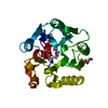
| ||||||||
|---|---|---|---|---|---|---|---|---|---|
| 1 |
| ||||||||
| Unit cell |
|
-Components
| #1: Protein | Mass: 32568.309 Da / Num. of mol.: 1 Source method: isolated from a genetically manipulated source Source: (gene. exp.) Homo sapiens (human) / Gene: DDAH1, DDAH / Plasmid: pQE30 / Production host:  Escherichia coli BL21(DE3) (bacteria) / Variant (production host): pREP4 / References: UniProt: O94760, dimethylargininase Escherichia coli BL21(DE3) (bacteria) / Variant (production host): pREP4 / References: UniProt: O94760, dimethylargininase | ||||
|---|---|---|---|---|---|
| #2: Chemical | ChemComp-M3B / (  Mass: 190.286 Da / Num. of mol.: 1 / Source method: obtained synthetically / Formula: C8H22N4O / Feature type: SUBJECT OF INVESTIGATION Mass: 190.286 Da / Num. of mol.: 1 / Source method: obtained synthetically / Formula: C8H22N4O / Feature type: SUBJECT OF INVESTIGATION | ||||
| #3: Chemical | Glycerol  Mass: 92.094 Da / Num. of mol.: 2 / Source method: obtained synthetically / Formula: C3H8O3 / Feature type: SUBJECT OF INVESTIGATION Mass: 92.094 Da / Num. of mol.: 2 / Source method: obtained synthetically / Formula: C3H8O3 / Feature type: SUBJECT OF INVESTIGATION#4: Water | ChemComp-HOH / | Water Mass: 18.015 Da / Num. of mol.: 137 / Source method: isolated from a natural source / Formula: H2O Mass: 18.015 Da / Num. of mol.: 137 / Source method: isolated from a natural source / Formula: H2OHas ligand of interest | Y | |
-Experimental details
-Experiment
| Experiment | Method: X-RAY DIFFRACTION / Number of used crystals: 1 |
|---|
- Sample preparation
| Crystal | Density Matthews: 2.12 Å3/Da / Density % sol: 42.08 % |
|---|---|
| Crystal grow | Temperature: 277.15 K / Method: vapor diffusion, sitting drop Details: reservoir: 0.1M MES pH 6.0 + 30% PEG6000 drop: 1+1 (v/v) cryo protectant: reservoir + 20%(v/v) glycerol |
-Data collection
| Diffraction | Mean temperature: 100 K / Serial crystal experiment: N |
|---|---|
| Diffraction source | Source: SYNCHROTRON / Site: PETRA III, DESY  / Beamline: P11 / Wavelength: 1.008 Å / Beamline: P11 / Wavelength: 1.008 Å |
| Detector | Type: DECTRIS PILATUS 6M / Detector: PIXEL / Date: Apr 4, 2013 |
| Radiation | Protocol: SINGLE WAVELENGTH / Monochromatic (M) / Laue (L): M / Scattering type: x-ray |
| Radiation wavelength | Wavelength: 1.008 Å / Relative weight: 1 |
| Reflection | Resolution: 1.76→46.56 Å / Num. obs: 25915 / % possible obs: 99.6 % / Redundancy: 6.67 % / CC1/2: 0.998 / Net I/σ(I): 11.98 |
| Reflection shell | Resolution: 1.76→1.8 Å / Mean I/σ(I) obs: 1.52 / Num. unique obs: 1824 / CC1/2: 0.675 / % possible all: 95.4 |
- Processing
| Software |
| ||||||||||||||||||||||||
|---|---|---|---|---|---|---|---|---|---|---|---|---|---|---|---|---|---|---|---|---|---|---|---|---|---|
| Refinement | Method to determine structure: MOLECULAR REPLACEMENT / Resolution: 1.76→46.56 Å / SU ML: 0.27 / Cross valid method: FREE R-VALUE / σ(F): 1.39 / Phase error: 24.95
| ||||||||||||||||||||||||
| Solvent computation | Shrinkage radii: 0.9 Å / VDW probe radii: 1.11 Å | ||||||||||||||||||||||||
| Displacement parameters | Biso max: 90.37 Å2 / Biso mean: 36.0639 Å2 / Biso min: 17.49 Å2 | ||||||||||||||||||||||||
| Refinement step | Cycle: final / Resolution: 1.76→46.56 Å
| ||||||||||||||||||||||||
| LS refinement shell | Resolution: 1.76→1.8 Å / Rfactor Rfree error: 0
|
