 Movie
Movie Controller

[English] 日本語
 Yorodumi
- PDB-6spo: Structure of the Escherichia coli methionyl-tRNA synthetase compl... -
+ Open data

- Basic information
| Entry | Database: PDB / ID: 6spo | ||||||
|---|---|---|---|---|---|---|---|
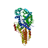
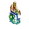
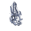
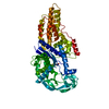
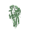
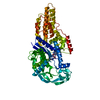
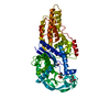
| Title | Structure of the Escherichia coli methionyl-tRNA synthetase complexed with methionine | ||||||
 Components Components | Methionine--tRNA ligase | ||||||
 Keywords Keywords |  TRANSLATION / tRNA aminoacylation TRANSLATION / tRNA aminoacylation | ||||||
| Function / homology |  Function and homology informationmethionine-tRNA ligase / methionine-tRNA ligase activity / methionyl-tRNA aminoacylation / tRNA binding / protein homodimerization activity / zinc ion binding / ATP binding / membrane / metal ion binding / cytosol / cytoplasm Function and homology informationmethionine-tRNA ligase / methionine-tRNA ligase activity / methionyl-tRNA aminoacylation / tRNA binding / protein homodimerization activity / zinc ion binding / ATP binding / membrane / metal ion binding / cytosol / cytoplasmSimilarity search - Function | ||||||
| Biological species |  Escherichia coli (E. coli) Escherichia coli (E. coli) | ||||||
| Method | X-RAY DIFFRACTION / SYNCHROTRON / MOLECULAR REPLACEMENT / Resolution: 1.2 Å | ||||||
 Authors Authors | Nigro, G. / Schmitt, E. / Mechulam, Y. | ||||||
 Citation Citation | Journal: J.Struct.Biol. / Year: 2020 Title: Use of beta3-methionine as an amino acid substrate of Escherichia coli methionyl-tRNA synthetase. Authors: Nigro, G. / Bourcier, S. / Lazennec-Schurdevin, C. / Schmitt, E. / Marliere, P. / Mechulam, Y. | ||||||
| History |
|
- Structure visualization
| Structure viewer | Molecule: MolmilJmol/JSmol |
|---|
- Downloads & links
-Download
| PDBx/mmCIF format | 6spo.cif.gz | 453.5 KB | Display | PDBx/mmCIF format |
|---|---|---|---|---|
| PDB format | pdb6spo.ent.gz | 307.7 KB | Display | PDB format |
| PDBx/mmJSON format | 6spo.json.gz | Tree view | PDBx/mmJSON format | |
| Others |  Other downloads Other downloads |
-Validation report
| Arichive directory | https://data.pdbj.org/pub/pdb/validation_reports/sp/6spoftp://data.pdbj.org/pub/pdb/validation_reports/sp/6spo | HTTPS FTP |
|---|
-Related structure data
| Related structure data |  6spnC 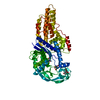 6sppC  6spqC  6sprC  1qqtS S: Starting model for refinement C: citing same article ( |
|---|---|
| Similar structure data |


-Links
 PDBj
PDBj
- Assembly
| Deposited unit | 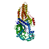
| ||||||||||||
|---|---|---|---|---|---|---|---|---|---|---|---|---|---|
| 1 |
| ||||||||||||
| Unit cell |
|
-Components
-Protein , 1 types, 1 molecules A
| #1: Protein | Mass: 64730.008 Da / Num. of mol.: 1 / Mutation: Truncated after residue 547; His-tagged Source method: isolated from a genetically manipulated source Source: (gene. exp.) Escherichia coli (E. coli)Gene: metG, BvCmsKSP058_01266, BvCmsNSP007_01600, ED648_21370, UN91_27160 Production host: Escherichia coli (E. coli)References: UniProt: A0A0F3U9S7, UniProt: P00959*PLUS, methionine-tRNA ligase |
|---|
-Non-polymers , 5 types, 752 molecules 


| #2: Chemical | ChemComp-ZN /  Mass: 65.409 Da / Num. of mol.: 1 / Source method: obtained synthetically / Formula: Zn Mass: 65.409 Da / Num. of mol.: 1 / Source method: obtained synthetically / Formula: Zn | ||||
|---|---|---|---|---|---|
| #3: Chemical | ChemComp-CIT / Citric acid Mass: 192.124 Da / Num. of mol.: 1 / Source method: obtained synthetically / Formula: C6H8O7 Mass: 192.124 Da / Num. of mol.: 1 / Source method: obtained synthetically / Formula: C6H8O7 | ||||
| #4: Chemical | Glycerol Mass: 92.094 Da / Num. of mol.: 3 / Source method: obtained synthetically / Formula: C3H8O3 Mass: 92.094 Da / Num. of mol.: 3 / Source method: obtained synthetically / Formula: C3H8O3#5: Chemical | ChemComp-MET / | Methionine Type: L-peptide linking / Mass: 149.211 Da / Num. of mol.: 1 / Source method: obtained synthetically / Formula: C5H11NO2S / Feature type: SUBJECT OF INVESTIGATION Type: L-peptide linking / Mass: 149.211 Da / Num. of mol.: 1 / Source method: obtained synthetically / Formula: C5H11NO2S / Feature type: SUBJECT OF INVESTIGATION#6: Water | ChemComp-HOH / | WaterMass: 18.015 Da / Num. of mol.: 746 / Source method: isolated from a natural source / Formula: H2O |
-Details
| Has ligand of interest | Y |
|---|
-Experimental details
-Experiment
| Experiment | Method: X-RAY DIFFRACTION / Number of used crystals: 1 |
|---|
- Sample preparation
| Crystal | Density Matthews: 2.33 Å3/Da / Density % sol: 47.28 % |
|---|---|
| Crystal grow | Temperature: 281 K / Method: vapor diffusion / pH: 7 Details: 1.08 M ammonium citrate, 10 mM potassium phosphate, pH 7.0 |
-Data collection
| Diffraction | Mean temperature: 100 K / Serial crystal experiment: N |
|---|---|
| Diffraction source | Source: SYNCHROTRON / Site: SOLEIL  / Beamline: PROXIMA 1 / Wavelength: 0.978 Å / Beamline: PROXIMA 1 / Wavelength: 0.978 Å |
| Detector | Type: DECTRIS PILATUS 6M / Detector: PIXEL / Date: Apr 7, 2018 |
| Radiation | Monochromator: CRYSTAL / Protocol: SINGLE WAVELENGTH / Monochromatic (M) / Laue (L): M / Scattering type: x-ray |
| Radiation wavelength | Wavelength: 0.978 Å / Relative weight: 1 |
| Reflection | Resolution: 1.2→33.1 Å / Num. obs: 178894 / % possible obs: 99 % / Redundancy: 6.4 % / Biso Wilson estimate: 13.44 Å2 / CC1/2: 0.998 / Rsym value: 0.105 / Net I/σ(I): 10 |
| Reflection shell | Resolution: 1.2→1.27 Å / Mean I/σ(I) obs: 1.11 / Num. unique obs: 29243 / CC1/2: 0.326 / Rsym value: 1.638 / % possible all: 94.8 |
- Processing
| Software |
| |||||||||||||||||||||||||||||||||||||||||||||||||||||||||||||||||||||||||||||||||||||||||||||||||||||||||||||||||||||||||||||||||||||||||||||||||||||||||||||||||||||||||||||||||||||||||||||||||||||||||||||||||||||||||
|---|---|---|---|---|---|---|---|---|---|---|---|---|---|---|---|---|---|---|---|---|---|---|---|---|---|---|---|---|---|---|---|---|---|---|---|---|---|---|---|---|---|---|---|---|---|---|---|---|---|---|---|---|---|---|---|---|---|---|---|---|---|---|---|---|---|---|---|---|---|---|---|---|---|---|---|---|---|---|---|---|---|---|---|---|---|---|---|---|---|---|---|---|---|---|---|---|---|---|---|---|---|---|---|---|---|---|---|---|---|---|---|---|---|---|---|---|---|---|---|---|---|---|---|---|---|---|---|---|---|---|---|---|---|---|---|---|---|---|---|---|---|---|---|---|---|---|---|---|---|---|---|---|---|---|---|---|---|---|---|---|---|---|---|---|---|---|---|---|---|---|---|---|---|---|---|---|---|---|---|---|---|---|---|---|---|---|---|---|---|---|---|---|---|---|---|---|---|---|---|---|---|---|---|---|---|---|---|---|---|---|---|---|---|---|---|---|---|---|
| Refinement | Method to determine structure: MOLECULAR REPLACEMENT Starting model: 1QQT Resolution: 1.2→33.05 Å / SU ML: 0.1557 / Cross valid method: FREE R-VALUE / σ(F): 1.33 / Phase error: 19.1559 Details: Refinement was performed using explicit riding hydrogen atoms generated in phenix.
| |||||||||||||||||||||||||||||||||||||||||||||||||||||||||||||||||||||||||||||||||||||||||||||||||||||||||||||||||||||||||||||||||||||||||||||||||||||||||||||||||||||||||||||||||||||||||||||||||||||||||||||||||||||||||
| Solvent computation | Shrinkage radii: 0.9 Å / VDW probe radii: 1.11 Å | |||||||||||||||||||||||||||||||||||||||||||||||||||||||||||||||||||||||||||||||||||||||||||||||||||||||||||||||||||||||||||||||||||||||||||||||||||||||||||||||||||||||||||||||||||||||||||||||||||||||||||||||||||||||||
| Displacement parameters | Biso mean: 19.25 Å2 | |||||||||||||||||||||||||||||||||||||||||||||||||||||||||||||||||||||||||||||||||||||||||||||||||||||||||||||||||||||||||||||||||||||||||||||||||||||||||||||||||||||||||||||||||||||||||||||||||||||||||||||||||||||||||
| Refinement step | Cycle: LAST / Resolution: 1.2→33.05 Å
| |||||||||||||||||||||||||||||||||||||||||||||||||||||||||||||||||||||||||||||||||||||||||||||||||||||||||||||||||||||||||||||||||||||||||||||||||||||||||||||||||||||||||||||||||||||||||||||||||||||||||||||||||||||||||
| Refine LS restraints |
| |||||||||||||||||||||||||||||||||||||||||||||||||||||||||||||||||||||||||||||||||||||||||||||||||||||||||||||||||||||||||||||||||||||||||||||||||||||||||||||||||||||||||||||||||||||||||||||||||||||||||||||||||||||||||
| LS refinement shell |
|
