 Movie
Movie Controller

[English] 日本語
 Yorodumi
- PDB-6rvo: Multicrystal dataset of thaumatin collected using a multilayer mo... -
+ Open data

- Basic information
| Entry | Database: PDB / ID: 6rvo | ||||||
|---|---|---|---|---|---|---|---|
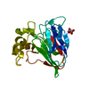
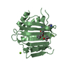
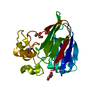
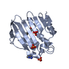
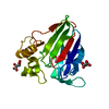
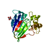
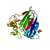
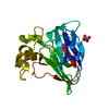
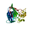
| Title | Multicrystal dataset of thaumatin collected using a multilayer monochromator. | ||||||
 Components Components | Thaumatin I | ||||||
 Keywords Keywords | PLANT PROTEIN / multilayer multicrystal VMXi | ||||||
| Function / homology |  Function and homology information Function and homology information | ||||||
| Biological species |  Thaumatococcus daniellii (katemfe) Thaumatococcus daniellii (katemfe) | ||||||
| Method | X-RAY DIFFRACTION / SYNCHROTRON / MOLECULAR REPLACEMENT / Resolution: 1.97 Å | ||||||
 Authors Authors | Sandy, J. / Sanchez-Weatherby, J. / Mikolajek, H. / Winter, G. | ||||||
 Citation Citation | Journal: Iucrj / Year: 2023 Title: Protein-to-structure pipeline for ambient-temperature crystallography at VMXi Authors: Mikolajek, H. / Sanchez-Weatherby, J. / Sandy, J. / Gildea, R.G. / Campeotto, I. / Cheruvara, H. / Clarke, J.D. / Foster, T. / Fujii, S. / Paulsen, I.T. / Shah, B.S. / Hough, M.A. | ||||||
| History |
|
- Structure visualization
| Structure viewer | Molecule: MolmilJmol/JSmol |
|---|
- Downloads & links
-Download
| PDBx/mmCIF format | 6rvo.cif.gz | 86.8 KB | Display | PDBx/mmCIF format |
|---|---|---|---|---|
| PDB format | pdb6rvo.ent.gz | 66.1 KB | Display | PDB format |
| PDBx/mmJSON format | 6rvo.json.gz | Tree view | PDBx/mmJSON format | |
| Others |  Other downloads Other downloads |
-Validation report
| Arichive directory | https://data.pdbj.org/pub/pdb/validation_reports/rv/6rvoftp://data.pdbj.org/pub/pdb/validation_reports/rv/6rvo | HTTPS FTP |
|---|
-Related structure data
| Related structure data |  6rzpC  6selC 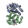 6svaC 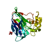 1rqwS S: Starting model for refinement C: citing same article ( |
|---|---|
| Similar structure data | |
| Experimental dataset #1 | Data reference: 10.5281/zenodo.3236085 / Data set type: diffraction image data |

-Links
 PDBj
PDBj

- Assembly
| Deposited unit | 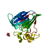
| ||||||||
|---|---|---|---|---|---|---|---|---|---|
| 1 |
| ||||||||
| Unit cell |
|
-Components
| #1: Protein | / Thaumatin-1 Mass: 22228.043 Da / Num. of mol.: 1 Source method: isolated from a genetically manipulated source Source: (gene. exp.) Thaumatococcus daniellii (katemfe) / Production host: Thaumatococcus daniellii (katemfe) / References: UniProt: P02883 |
|---|---|
| #2: Chemical | ChemComp-TLA / Tartaric acid  Mass: 150.087 Da / Num. of mol.: 1 / Source method: obtained synthetically / Formula: C4H6O6 Mass: 150.087 Da / Num. of mol.: 1 / Source method: obtained synthetically / Formula: C4H6O6 |
| #3: Water | ChemComp-HOH / Water Mass: 18.015 Da / Num. of mol.: 143 / Source method: isolated from a natural source / Formula: H2O Mass: 18.015 Da / Num. of mol.: 143 / Source method: isolated from a natural source / Formula: H2O |
-Experimental details
-Experiment
| Experiment | Method: X-RAY DIFFRACTION / Number of used crystals: 16 |
|---|
- Sample preparation
| Crystal | Density Matthews: 2.92 Å3/Da / Density % sol: 57.9 % |
|---|---|
| Crystal grow | Temperature: 293 K / Method: counter-diffusion / pH: 5.6 / Details: 0.1 M Sodium citrate pH 5.6 0.75M Na/K Tartrate |
-Data collection
| Diffraction | Mean temperature: 293 K / Serial crystal experiment: N | |||||||||||||||||||||||||||
|---|---|---|---|---|---|---|---|---|---|---|---|---|---|---|---|---|---|---|---|---|---|---|---|---|---|---|---|---|
| Diffraction source | Source: SYNCHROTRON / Site: Diamond  / Beamline: VMXi / Wavelength: 0.979 Å / Beamline: VMXi / Wavelength: 0.979 Å | |||||||||||||||||||||||||||
| Detector | Type: DECTRIS EIGER2 X 4M / Detector: PIXEL / Date: May 22, 2019 / Details: Multilayer monochromator | |||||||||||||||||||||||||||
| Radiation | Monochromator: Multilayer monochromator / Protocol: SINGLE WAVELENGTH / Monochromatic (M) / Laue (L): M / Scattering type: x-ray | |||||||||||||||||||||||||||
| Radiation wavelength | Wavelength: 0.979 Å / Relative weight: 1 | |||||||||||||||||||||||||||
| Reflection | Resolution: 1.97→58.56 Å / Num. obs: 19537 / % possible obs: 99.98 % / Redundancy: 38.7 % / CC1/2: 0.992 / Rmerge(I) obs: 0.409 / Rpim(I) all: 0.062 / Rrim(I) all: 0.414 / Net I/σ(I): 8.5 | |||||||||||||||||||||||||||
| Reflection shell | Diffraction-ID: 1
|
- Processing
| Software |
| |||||||||||||||||||||||||||||||||||||||||||||||||||||||||||||||||||||||||||||||||||||||||||||||||||||||||
|---|---|---|---|---|---|---|---|---|---|---|---|---|---|---|---|---|---|---|---|---|---|---|---|---|---|---|---|---|---|---|---|---|---|---|---|---|---|---|---|---|---|---|---|---|---|---|---|---|---|---|---|---|---|---|---|---|---|---|---|---|---|---|---|---|---|---|---|---|---|---|---|---|---|---|---|---|---|---|---|---|---|---|---|---|---|---|---|---|---|---|---|---|---|---|---|---|---|---|---|---|---|---|---|---|---|---|
| Refinement | Method to determine structure: MOLECULAR REPLACEMENT Starting model: 1RQW Resolution: 1.97→54.624 Å / SU ML: 0.19 / Cross valid method: FREE R-VALUE / σ(F): 1.34 / Phase error: 14.59 / Stereochemistry target values: ML
| |||||||||||||||||||||||||||||||||||||||||||||||||||||||||||||||||||||||||||||||||||||||||||||||||||||||||
| Solvent computation | Shrinkage radii: 0.9 Å / VDW probe radii: 1.11 Å / Solvent model: FLAT BULK SOLVENT MODEL | |||||||||||||||||||||||||||||||||||||||||||||||||||||||||||||||||||||||||||||||||||||||||||||||||||||||||
| Refinement step | Cycle: LAST / Resolution: 1.97→54.624 Å
| |||||||||||||||||||||||||||||||||||||||||||||||||||||||||||||||||||||||||||||||||||||||||||||||||||||||||
| Refine LS restraints |
| |||||||||||||||||||||||||||||||||||||||||||||||||||||||||||||||||||||||||||||||||||||||||||||||||||||||||
| LS refinement shell |
|
