 Movie
Movie Controller

[English] 日本語
 Yorodumi
- PDB-6rsa: NUCLEAR MAGNETIC RESONANCE AND NEUTRON DIFFRACTION STUDIES OF THE... -
+ Open data

- Basic information
| Entry | Database: PDB / ID: 6rsa | ||||||
|---|---|---|---|---|---|---|---|
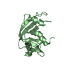
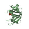
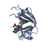
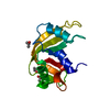
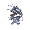
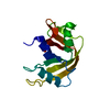
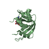
| Title | NUCLEAR MAGNETIC RESONANCE AND NEUTRON DIFFRACTION STUDIES OF THE COMPLEX OF RIBONUCLEASE*A WITH URIDINE VANADATE, A TRANSITION-STATE ANALOGUE | ||||||
 Components Components | RIBONUCLEASE A Pancreatic ribonuclease family Pancreatic ribonuclease family | ||||||
 Keywords Keywords | HYDROLASE | ||||||
| Function / homology |  Function and homology informationpancreatic ribonuclease / ribonuclease A activity / RNA nuclease activity / nucleic acid binding / lyase activity / defense response to Gram-positive bacterium / extracellular region Function and homology informationpancreatic ribonuclease / ribonuclease A activity / RNA nuclease activity / nucleic acid binding / lyase activity / defense response to Gram-positive bacterium / extracellular regionSimilarity search - Function | ||||||
| Biological species |  Bos taurus (cattle) Bos taurus (cattle) | ||||||
| Method | NEUTRON DIFFRACTION / SOLUTION NMR / Resolution: 2 Å | ||||||
 Authors Authors | Wlodawer, A. | ||||||
 Citation Citation | Journal: Biochemistry / Year: 1985 Title: Nuclear magnetic resonance and neutron diffraction studies of the complex of ribonuclease A with uridine vanadate, a transition-state analogue. Authors: Borah, B. / Chen, C.W. / Egan, W. / Miller, M. / Wlodawer, A. / Cohen, J.S. #1: Journal: Acta Crystallogr.,Sect.B / Year: 1986Title: Comparison of Two Independently Refined Models of Ribonuclease-A Authors: Wlodawer, A. / Borkakoti, N. / Moss, D.S. / Howlin, B. #2: Journal: Biochemistry / Year: 1983Title: Structure of Ribonuclease A. Results of Joint Neutron and X-Ray Refinement at 2.0-Angstroms Resolution Authors: Wlodawer, A. / Sjolin, L. #3: Journal: Proc.Natl.Acad.Sci.USA / Year: 1983Title: Active Site of Rnase. Neutron Diffraction Study of a Complex with Uridine Vanadate, a Transition-State Analog Authors: Wlodawer, A. / Miller, M. / Sjolin, L. #4: Journal: J.Biol.Chem. / Year: 1982Title: The Refined Crystal Structure of Ribonuclease A at 2.0 Angstroms Resolution Authors: Wlodawer, A. / Bott, R. / Sjolin, L. #5: Journal: Proc.Natl.Acad.Sci.USA / Year: 1982Title: Hydrogen Exchange in Rnase A. Neutron Diffraction Study Authors: Wlodawer, A. / Sjolin, L. #6: Journal: Acta Crystallogr.,Sect.A (Supplement) / Year: 1981Title: Structure of Ribonuclease A. X-Ray and Neutron Refinement Authors: Wlodawer, A. / Bott, R. / Sjolin, L. #7: Journal: Acta Crystallogr.,Sect.A (Supplement) / Year: 1981Title: Joint Refinement of Macromolecular Structures with X-Ray and Neutron Single-Crystal Diffraction Data Authors: Wlodawer, A. / Hendrickson, W.A. #8: Journal: Proc.Natl.Acad.Sci.USA / Year: 1981Title: Orientation of Histidine Residues in Rnase A. Neutron Diffraction Study Authors: Wlodawer, A. / Sjolin, L. #9: Journal: Acta Crystallogr.,Sect.B / Year: 1980Title: Studies of Ribonuclease-A by X-Ray and Neutron Diffraction Authors: Wlodawer, A. | ||||||
| History |
| ||||||
| Remark 700 | SHEET THIS STRUCTURE CONTAINS TWO SHEETS. SHEET S1 COMPRISES THREE STRANDS. IN THE SECOND STRAND OF ...SHEET THIS STRUCTURE CONTAINS TWO SHEETS. SHEET S1 COMPRISES THREE STRANDS. IN THE SECOND STRAND OF SHEET S1, RESIDUES 88 AND 89 *BULGE OUT*. IN ORDER TO REPRESENT THIS BREAK IN STRAND 2, TWO SHEETS (S1A AND S1B) ARE DEFINED BELOW. STRANDS 1 AND 3 OF *SHEETS* S1A AND S1B ARE, THEREFORE, IDENTICAL AND STRAND 2 DIFFERS. SHEET S2 COMPRISES FOUR STRANDS. RESIDUE 120 DOES NOT PROPERLY BELONG IN STRAND 4 OF SHEET S2. IN ORDER TO REPRESENT THIS BREAK IN STRAND 4, TWO SHEETS (S2A AND S2B) ARE DEFINED BELOW. STRANDS 1,2,3 OF *SHEETS* S2A AND S2B ARE, THEREFORE, IDENTICAL AND STRAND 4 DIFFERS. |
- Structure visualization
| Structure viewer | Molecule: MolmilJmol/JSmol |
|---|
- Downloads & links
-Download
| PDBx/mmCIF format | 6rsa.cif.gz | 60 KB | Display | PDBx/mmCIF format |
|---|---|---|---|---|
| PDB format | pdb6rsa.ent.gz | 50.4 KB | Display | PDB format |
| PDBx/mmJSON format | 6rsa.json.gz | Tree view | PDBx/mmJSON format | |
| Others |  Other downloads Other downloads |
-Validation report
| Arichive directory | https://data.pdbj.org/pub/pdb/validation_reports/rs/6rsaftp://data.pdbj.org/pub/pdb/validation_reports/rs/6rsa | HTTPS FTP |
|---|
-Related structure data
| Similar structure data |
|---|


-Links
 PDBj
PDBj


- Assembly
| Deposited unit | 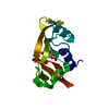
| |||||||||
|---|---|---|---|---|---|---|---|---|---|---|
| 1 |
| |||||||||
| Unit cell |
| |||||||||
| Atom site foot note | 1: RESIDUES 93 AND 114 ARE CIS-PROLINES. | |||||||||
| NMR ensembles |
|
-Components
| #1: Protein | Pancreatic ribonuclease family Mass: 13708.326 Da / Num. of mol.: 1 / Source method: isolated from a natural source / Source: (natural) Bos taurus (cattle) / Cell line: S2 / Organ: PANCREASReferences: UniProt: P00656, UniProt: P61823*PLUS, EC: 3.1.27.5 |
|---|---|
| #2: Chemical | ChemComp-UVC /   Mass: 343.141 Da / Num. of mol.: 1 / Source method: obtained synthetically / Formula: C9H12N2O9V Mass: 343.141 Da / Num. of mol.: 1 / Source method: obtained synthetically / Formula: C9H12N2O9V |
| #3: Chemical | ChemComp-DOD / Heavy water  Mass: 18.015 Da / Num. of mol.: 112 / Source method: isolated from a natural source / Formula: D2O Mass: 18.015 Da / Num. of mol.: 112 / Source method: isolated from a natural source / Formula: D2O |
| Nonpolymer details | COORDINATE |
-Experimental details
-Experiment
| Experiment |
|
|---|
- Sample preparation
| Crystal | Density Matthews: 2.18 Å3/Da / Density % sol: 43.65 % Description: DATA WERE COLLECTED ON DEUTERATED CRYSTALS. THE SOLVENT IS TERTIARY BUTANOL. AMIDE HYDROGENS WHICH EXCHANGED LESS THAN 50 PER CENT ARE ENTERED AS H, OTHERS ARE ENTERED AS D. |
|---|---|
| Crystal grow | *PLUS pH: 5.3 / Method: unknownDetails: Wlodawer, A., (1983) Proc.Nat.Acad.Sci.USA, 80, 3628. |
| Components of the solutions | *PLUS Conc.: 43 % / Common name: t-butyl alcohol |
- Processing
| Refinement | Resolution: 2→10 Å | ||||||||||||||||||||||||||||||||||||||||||||||||||||||||||||
|---|---|---|---|---|---|---|---|---|---|---|---|---|---|---|---|---|---|---|---|---|---|---|---|---|---|---|---|---|---|---|---|---|---|---|---|---|---|---|---|---|---|---|---|---|---|---|---|---|---|---|---|---|---|---|---|---|---|---|---|---|---|
| Refine LS restraints |
| ||||||||||||||||||||||||||||||||||||||||||||||||||||||||||||
| NMR ensemble | Conformers submitted total number: 1 | ||||||||||||||||||||||||||||||||||||||||||||||||||||||||||||
| Refinement | *PLUS Rfactor obs: 0.188 | ||||||||||||||||||||||||||||||||||||||||||||||||||||||||||||
| Solvent computation | *PLUS | ||||||||||||||||||||||||||||||||||||||||||||||||||||||||||||
| Displacement parameters | *PLUS |
