[English] 日本語
Yorodumi
- PDB-6rar: Pmar-Lig_PreS3-Mn -

+
Open data


ID or keywords:

Loading...

-
Basic information

Entry
Database: PDB / ID: 6rar
TitlePmar-Lig_PreS3-Mn
Components
  • ATP-dependent DNA ligase
  • DNA (5'-D(*TP*TP*CP*CP*GP*AP*CP*AP*GP*TP*GP*GP*GP*GP*TP*CP*GP*CP*AP*AP*T)-3')
  • DNA (5'-D(P*CP*AP*CP*TP*AP*TP*CP*GP*GP*AP*A)-3')
  • DNA/RNA (5'-D(*AP*TP*TP*GP*CP*GP*AP*C)-R(P*(OMC))-D(P*C)-3')
Keywords
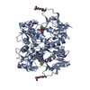
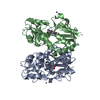
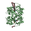
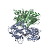
DNA BINDING PROTEIN / DNA ligase / ATP-dependent / ligase-DNA co-crystal structure / determinants in DNA binding
Function / homology
Function and homology information


DNA ligase (ATP) activity / DNA recombination / DNA replication / DNA repair / ATP binding
Similarity search - Function
DNA ligase, OB-like domain / DNA ligase OB-like domain / ATP-dependent DNA ligase AMP-binding site. / DNA ligase, ATP-dependent, conserved site / DNA ligase, ATP-dependent, central / ATP dependent DNA ligase domain / Nucleic acid-binding, OB-fold
Similarity search - Domain/homology
ADENOSINE MONOPHOSPHATE / : / DNA / DNA (> 10) / ATP-dependent DNA ligase
Similarity search - Component
Biological speciesProchlorococcus marinus str. MIT 9302 (bacteria)
synthetic construct (others)
MethodX-RAY DIFFRACTION / SYNCHROTRON / MOLECULAR REPLACEMENT / Resolution: 1.785 Å
AuthorsLeiros, H.K.S. / Williamson, A.
Funding support Norway, 1items
OrganizationGrant numberCountry
Research Council of Norway244247, 2015 Norway
CitationJournal: Nucleic Acids Res. / Year: 2019
Title: Structural intermediates of a DNA-ligase complex illuminate the role of the catalytic metal ion and mechanism of phosphodiester bond formation.
Authors: Williamson, A. / Leiros, H.S.
History
DepositionApr 7, 2019Deposition site: PDBE / Processing site: PDBE
Revision 1.0Jul 10, 2019Provider: repository / Type: Initial release
Revision 1.1Jan 29, 2020Group: Database references / Category: citation / citation_author
Item: _citation.country / _citation.journal_abbrev ..._citation.country / _citation.journal_abbrev / _citation.journal_id_ASTM / _citation.journal_id_CSD / _citation.journal_id_ISSN / _citation.journal_volume / _citation.page_first / _citation.page_last / _citation.pdbx_database_id_DOI / _citation.pdbx_database_id_PubMed / _citation.title / _citation.year / _citation_author.identifier_ORCID / _citation_author.name
Revision 1.2Jan 24, 2024Group: Data collection / Database references ...Data collection / Database references / Derived calculations / Refinement description
Category: chem_comp_atom / chem_comp_bond ...chem_comp_atom / chem_comp_bond / database_2 / pdbx_initial_refinement_model / pdbx_struct_conn_angle / struct_conn
Item: _database_2.pdbx_DOI / _database_2.pdbx_database_accession ..._database_2.pdbx_DOI / _database_2.pdbx_database_accession / _pdbx_struct_conn_angle.ptnr1_auth_seq_id / _pdbx_struct_conn_angle.ptnr3_auth_seq_id / _pdbx_struct_conn_angle.value / _struct_conn.conn_type_id / _struct_conn.id / _struct_conn.pdbx_dist_value / _struct_conn.pdbx_leaving_atom_flag / _struct_conn.ptnr1_auth_asym_id / _struct_conn.ptnr1_auth_comp_id / _struct_conn.ptnr1_auth_seq_id / _struct_conn.ptnr1_label_asym_id / _struct_conn.ptnr1_label_atom_id / _struct_conn.ptnr1_label_comp_id / _struct_conn.ptnr1_label_seq_id / _struct_conn.ptnr2_auth_asym_id / _struct_conn.ptnr2_auth_comp_id / _struct_conn.ptnr2_auth_seq_id / _struct_conn.ptnr2_label_asym_id / _struct_conn.ptnr2_label_atom_id / _struct_conn.ptnr2_label_comp_id / _struct_conn.ptnr2_label_seq_id

-
Structure visualization

Structure viewerMolecule:
MolmilJmol/JSmol

Downloads & links

-
Assembly

Deposited unit
B: DNA (5'-D(*TP*TP*CP*CP*GP*AP*CP*AP*GP*TP*GP*GP*GP*GP*TP*CP*GP*CP*AP*AP*T)-3')
C: DNA/RNA (5'-D(*AP*TP*TP*GP*CP*GP*AP*C)-R(P*(OMC))-D(P*C)-3')
D: DNA (5'-D(P*CP*AP*CP*TP*AP*TP*CP*GP*GP*AP*A)-3')
I: ATP-dependent DNA ligase
hetero molecules


Theoretical massNumber of molelcules
Total (without water)63,2417
Polymers62,7434
Non-polymers4983
Water7,170398
1


  • Idetical with deposited unit
  • defined by author&software
TypeNameSymmetry operationNumber
identity operation1_555x,y,z1
Buried area8950 Å2
ΔGint-80 kcal/mol
Surface area25240 Å2
MethodPISA
Unit cell
Length a, b, c (Å)67.304, 103.315, 68.543
Angle α, β, γ (deg.)90.00, 90.79, 90.00
Int Tables number4
Space group name H-MP1211

-
Components

-
DNA chain , 3 types, 3 molecules BCD

#1: DNA chain DNA (5'-D(*TP*TP*CP*CP*GP*AP*CP*AP*GP*TP*GP*GP*GP*GP*TP*CP*GP*CP*AP*AP*T)-3')


Mass: 6479.183 Da / Num. of mol.: 1 / Source method: obtained synthetically / Source: (synth.) synthetic construct (others)
#2: DNA chain DNA/RNA (5'-D(*AP*TP*TP*GP*CP*GP*AP*C)-R(P*(OMC))-D(P*C)-3')


Mass: 3035.007 Da / Num. of mol.: 1 / Source method: obtained synthetically / Source: (synth.) synthetic construct (others)
#3: DNA chain DNA (5'-D(P*CP*AP*CP*TP*AP*TP*CP*GP*GP*AP*A)-3')


Mass: 3342.212 Da / Num. of mol.: 1 / Source method: obtained synthetically / Source: (synth.) synthetic construct (others)

-
Protein , 1 types, 1 molecules I

#4: Protein ATP-dependent DNA ligase


Mass: 49886.480 Da / Num. of mol.: 1
Source method: isolated from a genetically manipulated source
Source: (gene. exp.) Prochlorococcus marinus str. MIT 9302 (bacteria)
Gene: EU96_0746 / Production host: Escherichia coli (E. coli) / References: UniProt: A0A0A2ACP7

-
Non-polymers , 4 types, 401 molecules

#5: Chemical ChemComp-MN / MANGANESE (II) ION


Mass: 54.938 Da / Num. of mol.: 1 / Source method: obtained synthetically / Formula: Mn
#6: Chemical ChemComp-AMP / ADENOSINE MONOPHOSPHATE / Adenosine monophosphate


Mass: 347.221 Da / Num. of mol.: 1 / Source method: obtained synthetically / Formula: C10H14N5O7P / Comment: AMP*YM
#7: Chemical ChemComp-SO4 / SULFATE ION / Sulfate


Mass: 96.063 Da / Num. of mol.: 1 / Source method: obtained synthetically / Formula: SO4
#8: Water ChemComp-HOH / water / Water


Mass: 18.015 Da / Num. of mol.: 398 / Source method: isolated from a natural source / Formula: H2O

-
Experimental details

-
Experiment

ExperimentMethod: X-RAY DIFFRACTION / Number of used crystals: 1

-
Sample preparation

CrystalDensity Matthews: 3.73 Å3/Da / Density % sol: 67.03 %
Crystal growTemperature: 277 K / Method: vapor diffusion
Details: 17-18% PEG 8K, 100 mM ammonium sulfate, 100 mM Bis-Tris pH 5.5.

-
Data collection

DiffractionMean temperature: 100 K / Serial crystal experiment: N
Diffraction sourceSource: SYNCHROTRON / Site: ESRF / Beamline: ID23-1 / Wavelength: 0.991872 Å
DetectorType: DECTRIS PILATUS3 S 6M / Detector: PIXEL / Date: Aug 25, 2018
RadiationProtocol: SINGLE WAVELENGTH / Monochromatic (M) / Laue (L): M / Scattering type: x-ray
Radiation wavelengthWavelength: 0.991872 Å / Relative weight: 1
ReflectionResolution: 1.785→68 Å / Num. obs: 57936 / % possible obs: 88.6 % / Redundancy: 3.1 % / Biso Wilson estimate: 29.1 Å2 / CC1/2: 0.993 / Rmerge(I) obs: 0.096 / Rpim(I) all: 0.084 / Net I/σ(I): 3.1
Reflection shellResolution: 1.785→1.98 Å / Rmerge(I) obs: 0.56 / Mean I/σ(I) obs: 2.4 / Num. unique obs: 2897 / CC1/2: 0.604 / Rpim(I) all: 0.425 / % possible all: 59.7

-
Processing

Software
NameVersionClassification
PHENIX(1.14_3260: ???)refinement
XDSdata reduction
Aimlessdata scaling
PHASERphasing
RefinementMethod to determine structure: MOLECULAR REPLACEMENT
Starting model: 6rau
Resolution: 1.785→24.291 Å / SU ML: 0.28 / Cross valid method: FREE R-VALUE / σ(F): 1.34 / Phase error: 37.43
RfactorNum. reflection% reflection
Rfree0.2639 2875 4.97 %
Rwork0.2325 --
obs0.2341 57870 88.6 %
Solvent computationShrinkage radii: 0.9 Å / VDW probe radii: 1.11 Å
Refinement stepCycle: LAST / Resolution: 1.785→24.291 Å
ProteinNucleic acidLigandSolventTotal
Num. atoms3433 856 28 398 4715
Refine LS restraints
Refine-IDTypeDev idealNumber
X-RAY DIFFRACTIONf_bond_d0.0184583
X-RAY DIFFRACTIONf_angle_d1.186341
X-RAY DIFFRACTIONf_dihedral_angle_d11.5123513
X-RAY DIFFRACTIONf_chiral_restr0.055686
X-RAY DIFFRACTIONf_plane_restr0.005663
LS refinement shell
Resolution (Å)Rfactor RfreeNum. reflection RfreeRfactor RworkNum. reflection RworkRefine-ID% reflection obs (%)
1.785-1.81420.3855100.358888X-RAY DIFFRACTION2
1.8142-1.84550.360280.3578198X-RAY DIFFRACTION5
1.8455-1.87910.4421120.3385346X-RAY DIFFRACTION9
1.8791-1.91520.3778230.314588X-RAY DIFFRACTION15
1.9152-1.95430.3401450.3055953X-RAY DIFFRACTION24
1.9543-1.99670.2991560.31031290X-RAY DIFFRACTION32
1.9967-2.04320.3344790.29091552X-RAY DIFFRACTION39
2.0432-2.09420.3099860.30141933X-RAY DIFFRACTION48
2.0942-2.15080.29951640.29012553X-RAY DIFFRACTION64
2.1508-2.21410.28191390.27313126X-RAY DIFFRACTION77
2.2141-2.28550.30421840.27373403X-RAY DIFFRACTION85
2.2855-2.36710.31581590.26053781X-RAY DIFFRACTION94
2.3671-2.46180.30242630.26293881X-RAY DIFFRACTION98
2.4618-2.57370.32192430.26433904X-RAY DIFFRACTION98
2.5737-2.70920.27891510.25923954X-RAY DIFFRACTION97
2.7092-2.87870.29492000.26633937X-RAY DIFFRACTION98
2.8787-3.10060.311990.25773944X-RAY DIFFRACTION98
3.1006-3.41180.26141850.22393873X-RAY DIFFRACTION96
3.4118-3.90380.24651910.20383845X-RAY DIFFRACTION95
3.9038-4.91170.21612340.18473923X-RAY DIFFRACTION97
4.9117-24.29350.23672440.21733923X-RAY DIFFRACTION96

+
About Yorodumi

-
News

-
Feb 9, 2022. New format data for meta-information of EMDB entries

New format data for meta-information of EMDB entries

  • Version 3 of the EMDB header file is now the official format.
  • The previous official version 1.9 will be removed from the archive.

Related info.:EMDB header

External links:wwPDB to switch to version 3 of the EMDB data model

-
Aug 12, 2020. Covid-19 info

Covid-19 info

URL: https://pdbj.org/emnavi/covid19.php

New page: Covid-19 featured information page in EM Navigator.

Related info.:Covid-19 info / Mar 5, 2020. Novel coronavirus structure data

+
Mar 5, 2020. Novel coronavirus structure data

Novel coronavirus structure data

Related info.:Yorodumi Speices / Aug 12, 2020. Covid-19 info

External links:COVID-19 featured content - PDBj / Molecule of the Month (242):Coronavirus Proteases

+
Jan 31, 2019. EMDB accession codes are about to change! (news from PDBe EMDB page)

EMDB accession codes are about to change! (news from PDBe EMDB page)

  • The allocation of 4 digits for EMDB accession codes will soon come to an end. Whilst these codes will remain in use, new EMDB accession codes will include an additional digit and will expand incrementally as the available range of codes is exhausted. The current 4-digit format prefixed with “EMD-” (i.e. EMD-XXXX) will advance to a 5-digit format (i.e. EMD-XXXXX), and so on. It is currently estimated that the 4-digit codes will be depleted around Spring 2019, at which point the 5-digit format will come into force.
  • The EM Navigator/Yorodumi systems omit the EMD- prefix.

Related info.:Q: What is EMD? / ID/Accession-code notation in Yorodumi/EM Navigator

External links:EMDB Accession Codes are Changing Soon! / Contact to PDBj

+
Jul 12, 2017. Major update of PDB

Major update of PDB

  • wwPDB released updated PDB data conforming to the new PDBx/mmCIF dictionary.
  • This is a major update changing the version number from 4 to 5, and with Remediation, in which all the entries are updated.
  • In this update, many items about electron microscopy experimental information are reorganized (e.g. em_software).
  • Now, EM Navigator and Yorodumi are based on the updated data.

External links:wwPDB Remediation / Enriched Model Files Conforming to OneDep Data Standards Now Available in the PDB FTP Archive

-
Yorodumi

Thousand views of thousand structures

  • Yorodumi is a browser for structure data from EMDB, PDB, SASBDB, etc.
  • This page is also the successor to EM Navigator detail page, and also detail information page/front-end page for Omokage search.
  • The word "yorodu" (or yorozu) is an old Japanese word meaning "ten thousand". "mi" (miru) is to see.

Related info.:EMDB / PDB / SASBDB / Comparison of 3 databanks / Yorodumi Search / Aug 31, 2016. New EM Navigator & Yorodumi / Yorodumi Papers / Jmol/JSmol / Function and homology information / Changes in new EM Navigator and Yorodumi

Read more