Movie
Movie Controller


+ Open data

- Basic information
| Entry | Database: PDB / ID: 6r91 | ||||||||||||||||||||||||
|---|---|---|---|---|---|---|---|---|---|---|---|---|---|---|---|---|---|---|---|---|---|---|---|---|---|
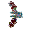
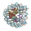
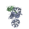
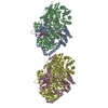
| Title | Cryo-EM structure of NCP_THF2(-3)-UV-DDB | ||||||||||||||||||||||||
 Components Components |
| ||||||||||||||||||||||||
 Keywords Keywords |  DNA BINDING PROTEIN / DNA damage / Nucleosome / 6-4 photoproduct DNA BINDING PROTEIN / DNA damage / Nucleosome / 6-4 photoproduct | ||||||||||||||||||||||||
| Function / homology |  Function and homology information Function and homology informationpositive regulation by virus of viral protein levels in host cell / epigenetic programming in the zygotic pronuclei / spindle assembly involved in female meiosis / Cul4-RING E3 ubiquitin ligase complex / UV-damage excision repair / biological process involved in interaction with symbiont / regulation of mitotic cell cycle phase transition / WD40-repeat domain binding / Cul4A-RING E3 ubiquitin ligase complex / Cul4B-RING E3 ubiquitin ligase complex ...positive regulation by virus of viral protein levels in host cell / epigenetic programming in the zygotic pronuclei / spindle assembly involved in female meiosis / Cul4-RING E3 ubiquitin ligase complex / UV-damage excision repair / biological process involved in interaction with symbiont / regulation of mitotic cell cycle phase transition / WD40-repeat domain binding / Cul4A-RING E3 ubiquitin ligase complex / Cul4B-RING E3 ubiquitin ligase complex / ubiquitin ligase complex scaffold activity / negative regulation of reproductive process / negative regulation of developmental process / site of DNA damage / cullin family protein binding / viral release from host cell / negative regulation of tumor necrosis factor-mediated signaling pathway / pyrimidine dimer repair / protein autoubiquitination / ectopic germ cell programmed cell death / negative regulation of megakaryocyte differentiation / positive regulation of viral genome replication / protein localization to CENP-A containing chromatin / Chromatin modifying enzymes / Replacement of protamines by nucleosomes in the male pronucleus / CENP-A containing nucleosome / epigenetic regulation of gene expression / Packaging Of Telomere Ends / response to UV / Recognition and association of DNA glycosylase with site containing an affected purine / Cleavage of the damaged purine / Deposition of new CENPA-containing nucleosomes at the centromere / positive regulation of gluconeogenesis / Recognition and association of DNA glycosylase with site containing an affected pyrimidine / Cleavage of the damaged pyrimidine / Inhibition of DNA recombination at telomere / Meiotic synapsis / telomere organization / RNA Polymerase I Promoter Opening / Interleukin-7 signaling / SUMOylation of chromatin organization proteins / Assembly of the ORC complex at the origin of replication / DNA methylation / Condensation of Prophase Chromosomes / HCMV Late Events / Chromatin modifications during the maternal to zygotic transition (MZT) / ERCC6 (CSB) and EHMT2 (G9a) positively regulate rRNA expression / SIRT1 negatively regulates rRNA expression / innate immune response in mucosa / PRC2 methylates histones and DNA / Defective pyroptosis / HDACs deacetylate histones / proteasomal protein catabolic process / Recognition of DNA damage by PCNA-containing replication complex / nucleotide-excision repair / RNA Polymerase I Promoter Escape / TP53 Regulates Transcription of DNA Repair Genes / Nonhomologous End-Joining (NHEJ) / lipopolysaccharide binding / Transcriptional regulation by small RNAs / Formation of the beta-catenin:TCF transactivating complex / DNA Damage Recognition in GG-NER / RUNX1 regulates genes involved in megakaryocyte differentiation and platelet function / Activated PKN1 stimulates transcription of AR (androgen receptor) regulated genes KLK2 and KLK3 / G2/M DNA damage checkpoint / NoRC negatively regulates rRNA expression / B-WICH complex positively regulates rRNA expression / Dual Incision in GG-NER / HDMs demethylate histones / regulation of circadian rhythm / DNA Damage/Telomere Stress Induced Senescence / Transcription-Coupled Nucleotide Excision Repair (TC-NER) / Metalloprotease DUBs / Formation of TC-NER Pre-Incision Complex / PKMTs methylate histone lysines / RMTs methylate histone arginines / Wnt signaling pathway / Meiotic recombination / Pre-NOTCH Transcription and Translation / Formation of Incision Complex in GG-NER / nucleosome assembly / protein polyubiquitination / Activation of anterior HOX genes in hindbrain development during early embryogenesis / HCMV Early Events / Dual incision in TC-NER / Gap-filling DNA repair synthesis and ligation in TC-NER / Transcriptional regulation of granulopoiesis / structural constituent of chromatin / positive regulation of protein catabolic process / UCH proteinases / cellular response to UV / rhythmic process / nucleosome / antimicrobial humoral immune response mediated by antimicrobial peptide / protein-macromolecule adaptor activity / E3 ubiquitin ligases ubiquitinate target proteins / site of double-strand break / cell junction / Recruitment and ATM-mediated phosphorylation of repair and signaling proteins at DNA double strand breaks / gene expressionSimilarity search - Function | ||||||||||||||||||||||||
| Biological species |  Homo sapiens (human) Homo sapiens (human) | ||||||||||||||||||||||||
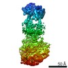
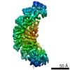
| Method | ELECTRON MICROSCOPY / single particle reconstruction / cryo EM / Resolution: 4.1 Å | ||||||||||||||||||||||||
 Authors Authors | Matsumoto, S. / Cavadini, S. / Bunker, R.D. / Thoma, N.H. | ||||||||||||||||||||||||
| Funding support |  Switzerland, Switzerland,  Japan, 7items Japan, 7items
| ||||||||||||||||||||||||
 Citation Citation | Journal: Nature / Year: 2019 Title: DNA damage detection in nucleosomes involves DNA register shifting. Authors: Syota Matsumoto / Simone Cavadini / Richard D Bunker / Ralph S Grand / Alessandro Potenza / Julius Rabl / Junpei Yamamoto / Andreas D Schenk / Dirk Schübeler / Shigenori Iwai / Kaoru ...Authors: Syota Matsumoto / Simone Cavadini / Richard D Bunker / Ralph S Grand / Alessandro Potenza / Julius Rabl / Junpei Yamamoto / Andreas D Schenk / Dirk Schübeler / Shigenori Iwai / Kaoru Sugasawa / Hitoshi Kurumizaka / Nicolas H Thomä / Abstract: Access to DNA packaged in nucleosomes is critical for gene regulation, DNA replication and DNA repair. In humans, the UV-damaged DNA-binding protein (UV-DDB) complex detects UV-light-induced ...Access to DNA packaged in nucleosomes is critical for gene regulation, DNA replication and DNA repair. In humans, the UV-damaged DNA-binding protein (UV-DDB) complex detects UV-light-induced pyrimidine dimers throughout the genome; however, it remains unknown how these lesions are recognized in chromatin, in which nucleosomes restrict access to DNA. Here we report cryo-electron microscopy structures of UV-DDB bound to nucleosomes bearing a 6-4 pyrimidine-pyrimidone dimer or a DNA-damage mimic in various positions. We find that UV-DDB binds UV-damaged nucleosomes at lesions located in the solvent-facing minor groove without affecting the overall nucleosome architecture. In the case of buried lesions that face the histone core, UV-DDB changes the predominant translational register of the nucleosome and selectively binds the lesion in an accessible, exposed position. Our findings explain how UV-DDB detects occluded lesions in strongly positioned nucleosomes, and identify slide-assisted site exposure as a mechanism by which high-affinity DNA-binding proteins can access otherwise occluded sites in nucleosomal DNA. | ||||||||||||||||||||||||
| History |
|
- Structure visualization
| Movie |
Movie viewer |
|---|---|
| Structure viewer | Molecule: MolmilJmol/JSmol |
- Downloads & links
-Download
| PDBx/mmCIF format | 6r91.cif.gz | 877.3 KB | Display | PDBx/mmCIF format |
|---|---|---|---|---|
| PDB format | pdb6r91.ent.gz | 716.9 KB | Display | PDB format |
| PDBx/mmJSON format | 6r91.json.gz | Tree view | PDBx/mmJSON format | |
| Others |  Other downloads Other downloads |
-Validation report
| Arichive directory | https://data.pdbj.org/pub/pdb/validation_reports/r9/6r91ftp://data.pdbj.org/pub/pdb/validation_reports/r9/6r91 | HTTPS FTP |
|---|
-Related structure data
| Related structure data | 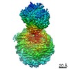 4765MC 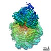 4762C 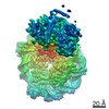 4763C 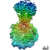 4764C 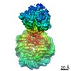 4766C 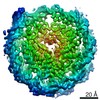 4767C 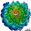 4768C 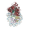 6r8yC  6r8zC 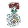 6r90C 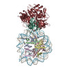 6r92C 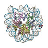 6r93C 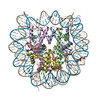 6r94C M: map data used to model this data C: citing same article ( |
|---|---|
| Similar structure data |


-Links
 PDBj
PDBj


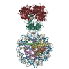
- Assembly
| Deposited unit | 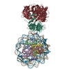
|
|---|---|
| 1 |
|
-Components
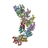
-Protein , 4 types, 8 molecules AEBFCGDH
| #1: Protein | Histone H3 / Histone H3/a / Histone H3/b / Histone H3/c / Histone H3/d / Histone H3/f / Histone H3/h / Histone ...Histone H3/a / Histone H3/b / Histone H3/c / Histone H3/d / Histone H3/f / Histone H3/h / Histone H3/i / Histone H3/j / Histone H3/k / Histone H3/l Mass: 15719.445 Da / Num. of mol.: 2 Source method: isolated from a genetically manipulated source Source: (gene. exp.) Homo sapiens (human)Gene: HIST1H3A, H3FA, HIST1H3B, H3FL, HIST1H3C, H3FC, HIST1H3D, H3FB, HIST1H3E, H3FD, HIST1H3F, H3FI, HIST1H3G, H3FH, HIST1H3H, H3FK, HIST1H3I, H3FF, HIST1H3J, H3FJ Production host:  Escherichia coli BL21(DE3) (bacteria) / References: UniProt: P68431 Escherichia coli BL21(DE3) (bacteria) / References: UniProt: P68431#2: Protein | Mass: 11676.703 Da / Num. of mol.: 2 Source method: isolated from a genetically manipulated source Source: (gene. exp.) Homo sapiens (human)Gene: HIST1H4A, H4/A, H4FA, HIST1H4B, H4/I, H4FI, HIST1H4C, H4/G, H4FG, HIST1H4D, H4/B, H4FB, HIST1H4E, H4/J, H4FJ, HIST1H4F, H4/C, H4FC, HIST1H4H, H4/H, H4FH, HIST1H4I, H4/M, H4FM, HIST1H4J, H4/E, ...Gene: HIST1H4A, H4/A, H4FA, HIST1H4B, H4/I, H4FI, HIST1H4C, H4/G, H4FG, HIST1H4D, H4/B, H4FB, HIST1H4E, H4/J, H4FJ, HIST1H4F, H4/C, H4FC, HIST1H4H, H4/H, H4FH, HIST1H4I, H4/M, H4FM, HIST1H4J, H4/E, H4FE, HIST1H4K, H4/D, H4FD, HIST1H4L, H4/K, H4FK, HIST2H4A, H4/N, H4F2, H4FN, HIST2H4, HIST2H4B, H4/O, H4FO, HIST4H4 Cell line (production host): JM109(DE3) / Production host: Escherichia coli (E. coli) / References: UniProt: P62805#3: Protein | Mass: 14447.825 Da / Num. of mol.: 2 Source method: isolated from a genetically manipulated source Source: (gene. exp.) Homo sapiens (human) / Gene: HIST1H2AB, H2AFM, HIST1H2AE, H2AFA / Production host: Escherichia coli BL21(DE3) (bacteria) / References: UniProt: P04908#4: Protein | Mass: 14217.516 Da / Num. of mol.: 2 Source method: isolated from a genetically manipulated source Source: (gene. exp.) Homo sapiens (human) / Gene: HIST1H2BJ, H2BFR / Production host: Escherichia coli BL21(DE3) (bacteria) / References: UniProt: P06899 |
|---|
-Human alpha-satellite DNA (145- ... , 2 types, 2 molecules IJ
| #5: DNA chain | Mass: 44756.648 Da / Num. of mol.: 1 / Source method: obtained synthetically / Source: (synth.) Homo sapiens (human) |
|---|---|
| #6: DNA chain | Mass: 44452.434 Da / Num. of mol.: 1 / Source method: obtained synthetically / Source: (synth.) Homo sapiens (human) |
-DNA damage-binding protein ... , 2 types, 2 molecules KL
| #7: Protein | Mass: 129766.305 Da / Num. of mol.: 1 Source method: isolated from a genetically manipulated source Source: (gene. exp.) Homo sapiens (human) / Gene: DDB1, XAP1 / Cell line (production host): High five / Production host:  Trichoplusia ni (cabbage looper) / References: UniProt: Q16531 Trichoplusia ni (cabbage looper) / References: UniProt: Q16531 |
|---|---|
| #8: Protein | Mass: 50601.844 Da / Num. of mol.: 1 Source method: isolated from a genetically manipulated source Source: (gene. exp.) Homo sapiens (human) / Gene: DDB2 / Cell line (production host): High five / Production host: Trichoplusia ni (cabbage looper) / References: UniProt: Q92466 |
-Experimental details
-Experiment
| Experiment | Method: ELECTRON MICROSCOPY |
|---|---|
| EM experiment | Aggregation state: PARTICLE / 3D reconstruction method: single particle reconstruction |
- Sample preparation
| Component |
| ||||||||||||||||||||||||||||||||||||
|---|---|---|---|---|---|---|---|---|---|---|---|---|---|---|---|---|---|---|---|---|---|---|---|---|---|---|---|---|---|---|---|---|---|---|---|---|---|
| Source (natural) |
| ||||||||||||||||||||||||||||||||||||
| Source (recombinant) |
| ||||||||||||||||||||||||||||||||||||
| Buffer solution | pH: 7.4 | ||||||||||||||||||||||||||||||||||||
| Specimen | Embedding applied: NO / Shadowing applied: NO / Staining applied: NO / Vitrification applied: YES | ||||||||||||||||||||||||||||||||||||
| Vitrification | Cryogen name: ETHANE / Humidity: 85 % / Chamber temperature: 277 K |
- Electron microscopy imaging
| Experimental equipment |  Model: Titan Krios / Image courtesy: FEI Company |
|---|---|
| Microscopy | Model: FEI TITAN KRIOS |
| Electron gun | Electron source: FIELD EMISSION GUN / Accelerating voltage: 300 kV / Illumination mode: SPOT SCAN |
| Electron lens | Mode: BRIGHT FIELDBright-field microscopy |
| Image recording | Electron dose: 40 e/Å2 / Film or detector model: GATAN K2 SUMMIT (4k x 4k) |
- Processing
| EM software |
| ||||||||||||||||||||||||||||||||||||||||||||||||||||
|---|---|---|---|---|---|---|---|---|---|---|---|---|---|---|---|---|---|---|---|---|---|---|---|---|---|---|---|---|---|---|---|---|---|---|---|---|---|---|---|---|---|---|---|---|---|---|---|---|---|---|---|---|---|
| CTF correction | Type: PHASE FLIPPING AND AMPLITUDE CORRECTION | ||||||||||||||||||||||||||||||||||||||||||||||||||||
| 3D reconstruction | Resolution: 4.1 Å / Resolution method: FSC 0.143 CUT-OFF / Num. of particles: 119309 / Symmetry type: POINT | ||||||||||||||||||||||||||||||||||||||||||||||||||||
| Atomic model building | Protocol: FLEXIBLE FIT / Space: REAL / Target criteria: Cross-correlation coefficient | ||||||||||||||||||||||||||||||||||||||||||||||||||||
| Atomic model building |
|