 Movie
Movie Controller


+ Open data

- Basic information
| Entry | Database: PDB / ID: 6r7v | ||||||
|---|---|---|---|---|---|---|---|
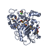
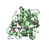
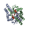
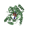
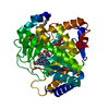
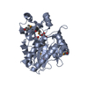
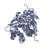
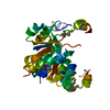
| Title | Tannerella forsythia promirolysin mutant E225A | ||||||
 Components Components | Mirolysin | ||||||
 Keywords Keywords |  HYDROLASE / metallopeptidase zymogen / metzincin / pappalysin family / Tannerella forsythia / periodontopathogen / periodontal disease HYDROLASE / metallopeptidase zymogen / metzincin / pappalysin family / Tannerella forsythia / periodontopathogen / periodontal disease | ||||||
| Function / homology | Peptidase M43, pregnancy-associated plasma-A / Pregnancy-associated plasma protein-A / Secretion system C-terminal sorting domain / Secretion system C-terminal sorting domain / Metallopeptidase, catalytic domain superfamily / metallopeptidase activity / metal ion binding / Mirolysin / Karilysin Function and homology information Function and homology information | ||||||
| Biological species |  Tannerella forsythia (bacteria) Tannerella forsythia (bacteria) | ||||||
| Method | X-RAY DIFFRACTION / SYNCHROTRON / MOLECULAR REPLACEMENT / Resolution: 1.4 Å | ||||||
 Authors Authors | Rodriguez-Banqueri, A. / Guevara, T. / Ksiazek, M. / Potempa, J. / Gomis-Ruth, F.X. | ||||||
 Citation Citation | Journal: Iucrj / Year: 2020 Title: Structure-based mechanism of cysteine-switch latency and of catalysis by pappalysin-family metallopeptidases. Authors: Guevara, T. / Rodriguez-Banqueri, A. / Ksiazek, M. / Potempa, J. / Gomis-Ruth, F.X. #1: Journal: Biol.Chem. / Year: 2017 Title: Mirolysin, a LysargiNase from Tannerella forsythia, proteolytically inactivates the human cathelicidin, LL-37. Authors: Koneru, L. / Ksiazek, M. / Waligorska, I. / Straczek, A. / Lukasik, M. / Madej, M. / Thogersen, I.B. / Enghild, J.J. / Potempa, J. | ||||||
| History |
|
- Structure visualization
| Structure viewer | Molecule: MolmilJmol/JSmol |
|---|
- Downloads & links
-Download
| PDBx/mmCIF format | 6r7v.cif.gz | 145 KB | Display | PDBx/mmCIF format |
|---|---|---|---|---|
| PDB format | pdb6r7v.ent.gz | 112.7 KB | Display | PDB format |
| PDBx/mmJSON format | 6r7v.json.gz | Tree view | PDBx/mmJSON format | |
| Others |  Other downloads Other downloads |
-Validation report
| Arichive directory | https://data.pdbj.org/pub/pdb/validation_reports/r7/6r7vftp://data.pdbj.org/pub/pdb/validation_reports/r7/6r7v | HTTPS FTP |
|---|
-Related structure data
| Related structure data |  6r7uSC  6r7wC S: Starting model for refinement C: citing same article ( |
|---|---|
| Similar structure data |


-Links
 PDBj
PDBj


- Assembly
| Deposited unit | 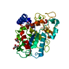
| ||||||||
|---|---|---|---|---|---|---|---|---|---|
| 1 |
| ||||||||
| Unit cell |
|
-Components
| #1: Protein | Mass: 35375.086 Da / Num. of mol.: 1 Source method: isolated from a genetically manipulated source Details: Extra N-terminal residues glycine-proline result from the cloning strategy. Source: (gene. exp.) Tannerella forsythia (bacteria) / Production host: Escherichia coli (E. coli) / References: UniProt: A0A0F7IPS1, UniProt: G8ULV1*PLUS | ||||||
|---|---|---|---|---|---|---|---|
| #2: Chemical |   Mass: 40.078 Da / Num. of mol.: 2 / Source method: obtained synthetically / Formula: Ca Mass: 40.078 Da / Num. of mol.: 2 / Source method: obtained synthetically / Formula: Ca#3: Chemical | ChemComp-ZN / |   Mass: 65.409 Da / Num. of mol.: 1 / Source method: obtained synthetically / Formula: Zn Mass: 65.409 Da / Num. of mol.: 1 / Source method: obtained synthetically / Formula: Zn#4: Chemical | ChemComp-GOL / Glycerol  Mass: 92.094 Da / Num. of mol.: 4 / Source method: obtained synthetically / Formula: C3H8O3 Mass: 92.094 Da / Num. of mol.: 4 / Source method: obtained synthetically / Formula: C3H8O3#5: Water | ChemComp-HOH / | Water Mass: 18.015 Da / Num. of mol.: 274 / Source method: isolated from a natural source / Formula: H2O Mass: 18.015 Da / Num. of mol.: 274 / Source method: isolated from a natural source / Formula: H2O |
-Experimental details
-Experiment
| Experiment | Method: X-RAY DIFFRACTION / Number of used crystals: 1 |
|---|
- Sample preparation
| Crystal | Density Matthews: 1.79 Å3/Da / Density % sol: 31.34 % |
|---|---|
| Crystal grow | Temperature: 293 K / Method: vapor diffusion, sitting drop Details: Promirolysin crystals were obtained at 20 degrees from drops containing 200 nL of protein solution at 0.6 mg/mL in 5 mM Tris-HCl pH 8.0, 50 mM sodium chloride and 100 nL of reservoir ...Details: Promirolysin crystals were obtained at 20 degrees from drops containing 200 nL of protein solution at 0.6 mg/mL in 5 mM Tris-HCl pH 8.0, 50 mM sodium chloride and 100 nL of reservoir solution, which comprised 25% PEG 1500, 0.1 M MIB buffer (malonic acid, imidazole and boric acid at a 2:3:3 molar ratio), pH 6.0. |
-Data collection
| Diffraction | Mean temperature: 100 K / Serial crystal experiment: N |
|---|---|
| Diffraction source | Source: SYNCHROTRON / Site: ALBA  / Beamline: XALOC / Wavelength: 1.2816 Å / Beamline: XALOC / Wavelength: 1.2816 Å |
| Detector | Type: DECTRIS PILATUS3 6M / Detector: PIXEL / Date: Nov 11, 2018 |
| Radiation | Protocol: SINGLE WAVELENGTH / Monochromatic (M) / Laue (L): M / Scattering type: x-ray |
| Radiation wavelength | Wavelength: 1.2816 Å / Relative weight: 1 |
| Reflection | Resolution: 1.4→51.4 Å / Num. obs: 50157 / % possible obs: 98.7 % / Redundancy: 9.7 % / Biso Wilson estimate: 19.77 Å2 / Net I/σ(I): 13.8 |
| Reflection shell | Resolution: 1.4→1.48 Å / Mean I/σ(I) obs: 1.8 / Num. unique obs: 7077 |
- Processing
| Software |
| |||||||||||||||||||||||||||||||||||||||||||||||||||||||||||||||||||||||||||||||||||||||||||||||||||||||||||||||||||||||||||||||||||||||||||||||||||||||||||||||||||||||||||||||
|---|---|---|---|---|---|---|---|---|---|---|---|---|---|---|---|---|---|---|---|---|---|---|---|---|---|---|---|---|---|---|---|---|---|---|---|---|---|---|---|---|---|---|---|---|---|---|---|---|---|---|---|---|---|---|---|---|---|---|---|---|---|---|---|---|---|---|---|---|---|---|---|---|---|---|---|---|---|---|---|---|---|---|---|---|---|---|---|---|---|---|---|---|---|---|---|---|---|---|---|---|---|---|---|---|---|---|---|---|---|---|---|---|---|---|---|---|---|---|---|---|---|---|---|---|---|---|---|---|---|---|---|---|---|---|---|---|---|---|---|---|---|---|---|---|---|---|---|---|---|---|---|---|---|---|---|---|---|---|---|---|---|---|---|---|---|---|---|---|---|---|---|---|---|---|---|---|
| Refinement | Method to determine structure: MOLECULAR REPLACEMENT Starting model: 6R7U Resolution: 1.4→20.34 Å / Cor.coef. Fo:Fc: 0.971 / Cor.coef. Fo:Fc free: 0.96 / SU R Cruickshank DPI: 0.06 / Cross valid method: THROUGHOUT / σ(F): 0 / SU R Blow DPI: 0.064 / SU Rfree Blow DPI: 0.065 / SU Rfree Cruickshank DPI: 0.062
| |||||||||||||||||||||||||||||||||||||||||||||||||||||||||||||||||||||||||||||||||||||||||||||||||||||||||||||||||||||||||||||||||||||||||||||||||||||||||||||||||||||||||||||||
| Displacement parameters | Biso mean: 25.37 Å2
| |||||||||||||||||||||||||||||||||||||||||||||||||||||||||||||||||||||||||||||||||||||||||||||||||||||||||||||||||||||||||||||||||||||||||||||||||||||||||||||||||||||||||||||||
| Refine analyze | Luzzati coordinate error obs: 0.16 Å | |||||||||||||||||||||||||||||||||||||||||||||||||||||||||||||||||||||||||||||||||||||||||||||||||||||||||||||||||||||||||||||||||||||||||||||||||||||||||||||||||||||||||||||||
| Refinement step | Cycle: 1 / Resolution: 1.4→20.34 Å
| |||||||||||||||||||||||||||||||||||||||||||||||||||||||||||||||||||||||||||||||||||||||||||||||||||||||||||||||||||||||||||||||||||||||||||||||||||||||||||||||||||||||||||||||
| Refine LS restraints |
| |||||||||||||||||||||||||||||||||||||||||||||||||||||||||||||||||||||||||||||||||||||||||||||||||||||||||||||||||||||||||||||||||||||||||||||||||||||||||||||||||||||||||||||||
| LS refinement shell | Resolution: 1.4→1.42 Å / Total num. of bins used: 36
| |||||||||||||||||||||||||||||||||||||||||||||||||||||||||||||||||||||||||||||||||||||||||||||||||||||||||||||||||||||||||||||||||||||||||||||||||||||||||||||||||||||||||||||||
| Refinement TLS params. | Method: refined / Refine-ID: X-RAY DIFFRACTION
| |||||||||||||||||||||||||||||||||||||||||||||||||||||||||||||||||||||||||||||||||||||||||||||||||||||||||||||||||||||||||||||||||||||||||||||||||||||||||||||||||||||||||||||||
| Refinement TLS group |
|
