 Movie
Movie Controller

[English] 日本語
 Yorodumi
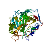
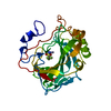
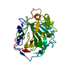
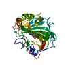
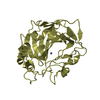
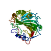
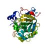
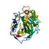
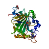
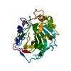
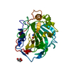
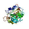
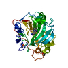
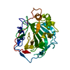
- PDB-6qfw: Human carbonic anhydrase II with bound IrCp* complex (cofactor 9)... -
+ Open data

- Basic information
| Entry | Database: PDB / ID: 6qfw | |||||||||||||||
|---|---|---|---|---|---|---|---|---|---|---|---|---|---|---|---|---|
| Title | Human carbonic anhydrase II with bound IrCp* complex (cofactor 9) to generate an artificial transfer hydrogenase (ATHase) | |||||||||||||||
 Components Components | Carbonic anhydrase 2 | |||||||||||||||
 Keywords Keywords | OXIDOREDUCTASE / Artificial Transfer Hydrogenase / bound IrCp* complex / zinc binding protein / human carbonic anhydrase II | |||||||||||||||
| Function / homology |  Function and homology information Function and homology informationpositive regulation of cellular pH reduction / positive regulation of dipeptide transmembrane transport / regulation of monoatomic anion transport / secretion / cyanamide hydratase / cyanamide hydratase activity / arylesterase activity / regulation of chloride transport / Reversible hydration of carbon dioxide / angiotensin-activated signaling pathway ...positive regulation of cellular pH reduction / positive regulation of dipeptide transmembrane transport / regulation of monoatomic anion transport / secretion / cyanamide hydratase / cyanamide hydratase activity / arylesterase activity / regulation of chloride transport / Reversible hydration of carbon dioxide / angiotensin-activated signaling pathway / positive regulation of synaptic transmission, GABAergic / regulation of intracellular pH / morphogenesis of an epithelium / carbonic anhydrase / carbonate dehydratase activity / carbon dioxide transport / Erythrocytes take up oxygen and release carbon dioxide / Erythrocytes take up carbon dioxide and release oxygen / neuron cellular homeostasis / one-carbon metabolic process / apical part of cell / myelin sheath / extracellular exosome / zinc ion binding / plasma membrane / cytosol / cytoplasmSimilarity search - Function | |||||||||||||||
| Biological species |  Homo sapiens (human) Homo sapiens (human) | |||||||||||||||
| Method | X-RAY DIFFRACTION / SYNCHROTRON / MOLECULAR REPLACEMENT / Resolution: 1.2 Å | |||||||||||||||
 Authors Authors | Rebelein, J.G. | |||||||||||||||
| Funding support |  Switzerland, Switzerland,  Germany, 4items Germany, 4items
| |||||||||||||||
 Citation Citation | Journal: Acs Catalysis / Year: 2019 Title: Chemical Optimization of Whole-Cell Transfer Hydrogenation Using Carbonic Anhydrase as Host Protein. Authors: Rebelein, J.G. / Cotelle, Y. / Garabedian, B. / Ward, T.R. | |||||||||||||||
| History |
|
- Structure visualization
| Structure viewer | Molecule: MolmilJmol/JSmol |
|---|
- Downloads & links
-Download
| PDBx/mmCIF format | 6qfw.cif.gz | 76.2 KB | Display | PDBx/mmCIF format |
|---|---|---|---|---|
| PDB format | pdb6qfw.ent.gz | 54.7 KB | Display | PDB format |
| PDBx/mmJSON format | 6qfw.json.gz | Tree view | PDBx/mmJSON format | |
| Others |  Other downloads Other downloads |
-Validation report
| Arichive directory | https://data.pdbj.org/pub/pdb/validation_reports/qf/6qfwftp://data.pdbj.org/pub/pdb/validation_reports/qf/6qfw | HTTPS FTP |
|---|
-Related structure data
| Related structure data | 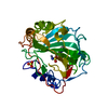 6qfuC 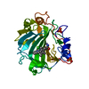 6qfvC  6qfxC 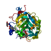 3zp9S S: Starting model for refinement C: citing same article ( |
|---|---|
| Similar structure data |


-Links
 PDBj
PDBj


- Assembly
| Deposited unit | 
| ||||||||
|---|---|---|---|---|---|---|---|---|---|
| 1 |
| ||||||||
| Unit cell |
|
-Components
-Protein , 1 types, 1 molecules A
| #1: Protein | / Carbonate dehydratase II / Carbonic anhydrase C / CAC / Carbonic anhydrase II / CA-II Mass: 29273.062 Da / Num. of mol.: 1 Source method: isolated from a genetically manipulated source Source: (gene. exp.) Homo sapiens (human) / Gene: CA2 / Production host:  Escherichia coli (E. coli) / References: UniProt: P00918, carbonic anhydrase Escherichia coli (E. coli) / References: UniProt: P00918, carbonic anhydrase |
|---|
-Non-polymers , 5 types, 271 molecules 


| #2: Chemical | ChemComp-JR8 /  Mass: 697.266 Da / Num. of mol.: 1 / Source method: obtained synthetically / Formula: C25H31ClIrN3O4S / Feature type: SUBJECT OF INVESTIGATION Mass: 697.266 Da / Num. of mol.: 1 / Source method: obtained synthetically / Formula: C25H31ClIrN3O4S / Feature type: SUBJECT OF INVESTIGATION |
|---|---|
| #3: Chemical | ChemComp-ZN /  Mass: 65.409 Da / Num. of mol.: 1 / Source method: obtained synthetically / Formula: Zn Mass: 65.409 Da / Num. of mol.: 1 / Source method: obtained synthetically / Formula: Zn |
| #4: Chemical | ChemComp-SO4 / Sulfate Mass: 96.063 Da / Num. of mol.: 1 / Source method: obtained synthetically / Formula: SO4 Mass: 96.063 Da / Num. of mol.: 1 / Source method: obtained synthetically / Formula: SO4 |
| #5: Chemical | ChemComp-IR /  Mass: 192.217 Da / Num. of mol.: 1 / Source method: obtained synthetically / Formula: Ir Mass: 192.217 Da / Num. of mol.: 1 / Source method: obtained synthetically / Formula: Ir |
| #6: Water | ChemComp-HOH / WaterMass: 18.015 Da / Num. of mol.: 267 / Source method: isolated from a natural source / Formula: H2O |
-Experimental details
-Experiment
| Experiment | Method: X-RAY DIFFRACTION / Number of used crystals: 1 |
|---|
- Sample preparation
| Crystal | Density Matthews: 2.13 Å3/Da / Density % sol: 42.26 % |
|---|---|
| Crystal grow | Temperature: 293 K / Method: vapor diffusion, sitting drop / pH: 7.9 / Details: 2.6 M ammonium sulfate, 50 mM Tris-H2SO4 |
-Data collection
| Diffraction | Mean temperature: 100 K / Serial crystal experiment: N |
|---|---|
| Diffraction source | Source: SYNCHROTRON / Site: SLS / Beamline: X06DA / Wavelength: 1 Å |
| Detector | Type: DECTRIS EIGER X 16M / Detector: PIXEL / Date: Nov 9, 2018 |
| Radiation | Protocol: SINGLE WAVELENGTH / Monochromatic (M) / Laue (L): M / Scattering type: x-ray |
| Radiation wavelength | Wavelength: 1 Å / Relative weight: 1 |
| Reflection | Resolution: 1.2→40.95 Å / Num. obs: 70789 / % possible obs: 98 % / Redundancy: 9.3 % / CC1/2: 0.995 / Rmerge(I) obs: 0.134 / Net I/σ(I): 13.5 |
| Reflection shell | Resolution: 1.2→1.23 Å / Redundancy: 6.4 % / Rmerge(I) obs: 0.2 / Mean I/σ(I) obs: 7 / Num. unique obs: 5020 / CC1/2: 0.975 / % possible all: 94.4 |
- Processing
| Software |
| |||||||||||||||||||||||||||||||||||||||||||||||||||||||||||||||||||||||||||
|---|---|---|---|---|---|---|---|---|---|---|---|---|---|---|---|---|---|---|---|---|---|---|---|---|---|---|---|---|---|---|---|---|---|---|---|---|---|---|---|---|---|---|---|---|---|---|---|---|---|---|---|---|---|---|---|---|---|---|---|---|---|---|---|---|---|---|---|---|---|---|---|---|---|---|---|---|
| Refinement | Method to determine structure: MOLECULAR REPLACEMENT Starting model: 3ZP9 Resolution: 1.2→40.95 Å / Cor.coef. Fo:Fc: 0.973 / Cor.coef. Fo:Fc free: 0.969 / Cross valid method: THROUGHOUT / σ(F): 0 / ESU R: 0.04 / ESU R Free: 0.041 / Stereochemistry target values: MAXIMUM LIKELIHOOD Details: HYDROGENS HAVE BEEN ADDED IN THE RIDING POSITIONS U VALUES : REFINED INDIVIDUALLY
| |||||||||||||||||||||||||||||||||||||||||||||||||||||||||||||||||||||||||||
| Solvent computation | Ion probe radii: 0.8 Å / Shrinkage radii: 0.8 Å / VDW probe radii: 1.2 Å / Solvent model: MASK | |||||||||||||||||||||||||||||||||||||||||||||||||||||||||||||||||||||||||||
| Displacement parameters | Biso max: 132.48 Å2 / Biso mean: 14.7 Å2 / Biso min: 5.77 Å2
| |||||||||||||||||||||||||||||||||||||||||||||||||||||||||||||||||||||||||||
| Refinement step | Cycle: final / Resolution: 1.2→40.95 Å
| |||||||||||||||||||||||||||||||||||||||||||||||||||||||||||||||||||||||||||
| Refine LS restraints |
| |||||||||||||||||||||||||||||||||||||||||||||||||||||||||||||||||||||||||||
| LS refinement shell | Resolution: 1.2→1.231 Å / Total num. of bins used: 20
|
