 Movie
Movie Controller

[English] 日本語
 Yorodumi
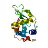
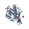
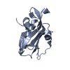
- PDB-6q10: Crystal structure of the soluble domain (residues 71-217) of a co... -
+ Open data

- Basic information
| Entry | Database: PDB / ID: 6q10 | ||||||
|---|---|---|---|---|---|---|---|
| Title | Crystal structure of the soluble domain (residues 71-217) of a conserved hypothetical secreted protein (Rv2700 ortholog) from Mycobacterium marinum | ||||||
 Components Components | MymaA.19257.a.B3 | ||||||
 Keywords Keywords | UNKNOWN FUNCTION / SSGCID / Mycobacerium marinum /  Structural Genomics / Seattle Structural Genomics Center for Infectious Disease / secreted protein / Rv2700 ortholog Structural Genomics / Seattle Structural Genomics Center for Infectious Disease / secreted protein / Rv2700 ortholog | ||||||
| Function / homology | LytR/CpsA/Psr regulator, C-terminal domain / LytR cell envelope-related transcriptional attenuator / membrane => GO:0016020 / Conserved hypothetical secreted protein Function and homology information Function and homology information | ||||||
| Biological species |  Mycobacterium marinum (bacteria) Mycobacterium marinum (bacteria) | ||||||
| Method | X-RAY DIFFRACTION / SYNCHROTRON / SAD / Resolution: 1.6 Å | ||||||
 Authors Authors | Seattle Structural Genomics Center for Infectious Disease (SSGCID) | ||||||
 Citation Citation | Journal: to be published Title: SSGCID, Mycobacerium marinum, Structural Genomics, Seattle Structural Genomics Center for Infectious Disease, secreted protein, Rv2700 ortholog Authors: Phan, J.N. / Abendroth, J. / Lorimer, D.D. / Horanyi, P.S. / Edwards, T.E. | ||||||
| History |
|
- Structure visualization
| Structure viewer | Molecule: MolmilJmol/JSmol |
|---|
- Downloads & links
-Download
| PDBx/mmCIF format | 6q10.cif.gz | 70.5 KB | Display | PDBx/mmCIF format |
|---|---|---|---|---|
| PDB format | pdb6q10.ent.gz | 54 KB | Display | PDB format |
| PDBx/mmJSON format | 6q10.json.gz | Tree view | PDBx/mmJSON format | |
| Others |  Other downloads Other downloads |
-Validation report
| Arichive directory | https://data.pdbj.org/pub/pdb/validation_reports/q1/6q10ftp://data.pdbj.org/pub/pdb/validation_reports/q1/6q10 | HTTPS FTP |
|---|
-Related structure data
| Similar structure data | |
|---|---|
| Other databases |


-Links
 PDBj
PDBj- Assembly
| Deposited unit | 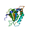
| ||||||||
|---|---|---|---|---|---|---|---|---|---|
| 1 |
| ||||||||
| Unit cell |
|
-Components
| #1: Protein | Mass: 16339.134 Da / Num. of mol.: 1 Source method: isolated from a genetically manipulated source Source: (gene. exp.) Mycobacterium marinum (strain ATCC BAA-535 / M) (bacteria)Strain: ATCC BAA-535 / M / Gene: MMAR_2014 Production host: Escherichia coli 'BL21-Gold(DE3)pLysS AG' (bacteria)Strain (production host): BL21(DE3) / References: UniProt: B2HM00 | ||||
|---|---|---|---|---|---|
| #2: Chemical | Ethylene glycol  Mass: 62.068 Da / Num. of mol.: 2 / Source method: obtained synthetically / Formula: C2H6O2 Mass: 62.068 Da / Num. of mol.: 2 / Source method: obtained synthetically / Formula: C2H6O2#3: Water | ChemComp-HOH / | Water Mass: 18.015 Da / Num. of mol.: 122 / Source method: isolated from a natural source / Formula: H2O Mass: 18.015 Da / Num. of mol.: 122 / Source method: isolated from a natural source / Formula: H2OHas ligand of interest | N | |
-Experimental details
-Experiment
| Experiment | Method: X-RAY DIFFRACTION / Number of used crystals: 1 |
|---|
- Sample preparation
| Crystal | Density Matthews: 2.1 Å3/Da / Density % sol: 41.5 % |
|---|---|
| Crystal grow | Temperature: 290 K / Method: vapor diffusion, sitting drop Details: Hampton Research Index screen, condition H1 (305884h1): 100mM Citric acid pH 3.5, 2000mM Ammonium sulfate: MymaA.19257.a.B3.PS38530 at 22.5mg/ml cryo: 25% EG: tray 305841 H1: puck bqx9-4. ...Details: Hampton Research Index screen, condition H1 (305884h1): 100mM Citric acid pH 3.5, 2000mM Ammonium sulfate: MymaA.19257.a.B3.PS38530 at 22.5mg/ml cryo: 25% EG: tray 305841 H1: puck bqx9-4. For experimental phasing, a crystal from the same condition, Index H1 (305884h1), was soaked for 15 seconds each in a mix of 87.5% reservoir / 12.5% 2.5M sodium iodide in ethylene glycol, followed by a soak in 75% reservoir / 25% 2.5M sodium iodide in ethylene glycol, vitrified in liquid nitrogen for in-house data collection at Cu-Kalpha wavelength: puck vgz4-3 |
-Data collection
| Diffraction |
| ||||||||||||||||||||||||||||||||||||||||||||||||||||||||||||||||||||||||||||||||||||||||||||||||||||||||||||||||||||||||||||||||||||||||||||||||||||||||||||||||||||||||||||||||||||||||||||||||||||||||||||||||||
|---|---|---|---|---|---|---|---|---|---|---|---|---|---|---|---|---|---|---|---|---|---|---|---|---|---|---|---|---|---|---|---|---|---|---|---|---|---|---|---|---|---|---|---|---|---|---|---|---|---|---|---|---|---|---|---|---|---|---|---|---|---|---|---|---|---|---|---|---|---|---|---|---|---|---|---|---|---|---|---|---|---|---|---|---|---|---|---|---|---|---|---|---|---|---|---|---|---|---|---|---|---|---|---|---|---|---|---|---|---|---|---|---|---|---|---|---|---|---|---|---|---|---|---|---|---|---|---|---|---|---|---|---|---|---|---|---|---|---|---|---|---|---|---|---|---|---|---|---|---|---|---|---|---|---|---|---|---|---|---|---|---|---|---|---|---|---|---|---|---|---|---|---|---|---|---|---|---|---|---|---|---|---|---|---|---|---|---|---|---|---|---|---|---|---|---|---|---|---|---|---|---|---|---|---|---|---|---|---|---|---|---|
| Diffraction source |
| ||||||||||||||||||||||||||||||||||||||||||||||||||||||||||||||||||||||||||||||||||||||||||||||||||||||||||||||||||||||||||||||||||||||||||||||||||||||||||||||||||||||||||||||||||||||||||||||||||||||||||||||||||
| Detector |
| ||||||||||||||||||||||||||||||||||||||||||||||||||||||||||||||||||||||||||||||||||||||||||||||||||||||||||||||||||||||||||||||||||||||||||||||||||||||||||||||||||||||||||||||||||||||||||||||||||||||||||||||||||
| Radiation |
| ||||||||||||||||||||||||||||||||||||||||||||||||||||||||||||||||||||||||||||||||||||||||||||||||||||||||||||||||||||||||||||||||||||||||||||||||||||||||||||||||||||||||||||||||||||||||||||||||||||||||||||||||||
| Radiation wavelength |
| ||||||||||||||||||||||||||||||||||||||||||||||||||||||||||||||||||||||||||||||||||||||||||||||||||||||||||||||||||||||||||||||||||||||||||||||||||||||||||||||||||||||||||||||||||||||||||||||||||||||||||||||||||
| Reflection | Resolution: 1.6→38.7 Å / Num. obs: 18638 / % possible obs: 99.1 % / Redundancy: 6.683 % / Biso Wilson estimate: 31.596 Å2 / CC1/2: 1 / Rmerge(I) obs: 0.029 / Rrim(I) all: 0.031 / Χ2: 1.03 / Net I/σ(I): 31.67 / Num. measured all: 124550 / Scaling rejects: 2 | ||||||||||||||||||||||||||||||||||||||||||||||||||||||||||||||||||||||||||||||||||||||||||||||||||||||||||||||||||||||||||||||||||||||||||||||||||||||||||||||||||||||||||||||||||||||||||||||||||||||||||||||||||
| Reflection shell | Diffraction-ID: 1
|

- Processing
| Software |
| ||||||||||||||||||||||||||||||||||||||||||||||||||||||||||||||||||||||||||||||||||||||||||||||||||||||||||||||||||||||||||||||||||||||||||||||||||||||
|---|---|---|---|---|---|---|---|---|---|---|---|---|---|---|---|---|---|---|---|---|---|---|---|---|---|---|---|---|---|---|---|---|---|---|---|---|---|---|---|---|---|---|---|---|---|---|---|---|---|---|---|---|---|---|---|---|---|---|---|---|---|---|---|---|---|---|---|---|---|---|---|---|---|---|---|---|---|---|---|---|---|---|---|---|---|---|---|---|---|---|---|---|---|---|---|---|---|---|---|---|---|---|---|---|---|---|---|---|---|---|---|---|---|---|---|---|---|---|---|---|---|---|---|---|---|---|---|---|---|---|---|---|---|---|---|---|---|---|---|---|---|---|---|---|---|---|---|---|---|---|---|
| Refinement | Method to determine structure: SAD / Resolution: 1.6→38.7 Å / SU ML: 0.15 / Cross valid method: THROUGHOUT / σ(F): 1.36 / Phase error: 19.25
| ||||||||||||||||||||||||||||||||||||||||||||||||||||||||||||||||||||||||||||||||||||||||||||||||||||||||||||||||||||||||||||||||||||||||||||||||||||||
| Solvent computation | Shrinkage radii: 0.9 Å / VDW probe radii: 1.11 Å | ||||||||||||||||||||||||||||||||||||||||||||||||||||||||||||||||||||||||||||||||||||||||||||||||||||||||||||||||||||||||||||||||||||||||||||||||||||||
| Displacement parameters | Biso max: 78.09 Å2 / Biso mean: 31.2634 Å2 / Biso min: 15.62 Å2 | ||||||||||||||||||||||||||||||||||||||||||||||||||||||||||||||||||||||||||||||||||||||||||||||||||||||||||||||||||||||||||||||||||||||||||||||||||||||
| Refinement step | Cycle: final / Resolution: 1.6→38.7 Å
| ||||||||||||||||||||||||||||||||||||||||||||||||||||||||||||||||||||||||||||||||||||||||||||||||||||||||||||||||||||||||||||||||||||||||||||||||||||||
| Refine LS restraints |
| ||||||||||||||||||||||||||||||||||||||||||||||||||||||||||||||||||||||||||||||||||||||||||||||||||||||||||||||||||||||||||||||||||||||||||||||||||||||
| LS refinement shell | Refine-ID: X-RAY DIFFRACTION / Rfactor Rfree error: 0
| ||||||||||||||||||||||||||||||||||||||||||||||||||||||||||||||||||||||||||||||||||||||||||||||||||||||||||||||||||||||||||||||||||||||||||||||||||||||
| Refinement TLS params. | Method: refined / Refine-ID: X-RAY DIFFRACTION
| ||||||||||||||||||||||||||||||||||||||||||||||||||||||||||||||||||||||||||||||||||||||||||||||||||||||||||||||||||||||||||||||||||||||||||||||||||||||
| Refinement TLS group |
|
