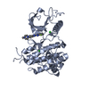
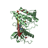
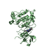
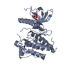
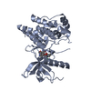
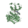
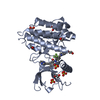
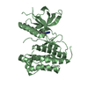
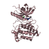
[English] 日本語
Yorodumi
- PDB-6p68: Crystal structure of FGFR1-Y563C (FGFR4 surrogate) covalently bou... -

+
Open data


ID or keywords:

Loading...

-
Basic information

Entry
Database: PDB / ID: 6p68
TitleCrystal structure of FGFR1-Y563C (FGFR4 surrogate) covalently bound to compound 22.
ComponentsFibroblast growth factor receptor 1
KeywordsTRANSFERASE / TRANSFERASE-TRANSFER complex
Function / homology
Function and homology information


Signaling by FGFR1 amplification mutants / negative regulation of fibroblast growth factor production / positive regulation of mitotic cell cycle DNA replication / regulation of extrinsic apoptotic signaling pathway in absence of ligand / Signaling by plasma membrane FGFR1 fusions / diphosphate metabolic process / FGFR1c and Klotho ligand binding and activation / vitamin D3 metabolic process / regulation of phosphate transport / regulation of lateral mesodermal cell fate specification ...Signaling by FGFR1 amplification mutants / negative regulation of fibroblast growth factor production / positive regulation of mitotic cell cycle DNA replication / regulation of extrinsic apoptotic signaling pathway in absence of ligand / Signaling by plasma membrane FGFR1 fusions / diphosphate metabolic process / FGFR1c and Klotho ligand binding and activation / vitamin D3 metabolic process / regulation of phosphate transport / regulation of lateral mesodermal cell fate specification / positive regulation of MAPKKK cascade by fibroblast growth factor receptor signaling pathway / cementum mineralization / positive regulation of endothelial cell chemotaxis to fibroblast growth factor / receptor-receptor interaction / response to sodium phosphate / regulation of branching involved in salivary gland morphogenesis by mesenchymal-epithelial signaling / fibroblast growth factor receptor signaling pathway involved in orbitofrontal cortex development / auditory receptor cell development / ventricular zone neuroblast division / Epithelial-Mesenchymal Transition (EMT) during gastrulation / positive regulation of parathyroid hormone secretion / chordate embryonic development / mesenchymal cell proliferation / paraxial mesoderm development / fibroblast growth factor receptor activity / FGFR1b ligand binding and activation / branching involved in salivary gland morphogenesis / Signaling by activated point mutants of FGFR1 / FGFR1c ligand binding and activation / organ induction / Downstream signaling of activated FGFR1 / Phospholipase C-mediated cascade: FGFR1 / positive regulation of phospholipase activity / lung-associated mesenchyme development / cell projection assembly / phosphatidylinositol-mediated signaling / outer ear morphogenesis / middle ear morphogenesis / embryonic limb morphogenesis / skeletal system morphogenesis / ureteric bud development / positive regulation of vascular endothelial cell proliferation / positive regulation of mesenchymal cell proliferation / cardiac muscle cell proliferation / inner ear morphogenesis / midbrain development / fibroblast growth factor binding / positive regulation of stem cell proliferation / Formation of paraxial mesoderm / regulation of cell differentiation / PI-3K cascade:FGFR1 / PI3K Cascade / epithelial to mesenchymal transition / positive regulation of blood vessel endothelial cell migration / fibroblast growth factor receptor signaling pathway / calcium ion homeostasis / chondrocyte differentiation / positive regulation of phospholipase C activity / SHC-mediated cascade:FGFR1 / cell maturation / positive regulation of cardiac muscle cell proliferation / FRS-mediated FGFR1 signaling / positive regulation of neuron differentiation / Signaling by FGFR1 in disease / SH2 domain binding / NCAM signaling for neurite out-growth / Signal transduction by L1 / skeletal system development / stem cell proliferation / stem cell differentiation / positive regulation of cell differentiation / sensory perception of sound / Negative regulation of FGFR1 signaling / neuron migration / positive regulation of MAP kinase activity / positive regulation of neuron projection development / receptor protein-tyrosine kinase / peptidyl-tyrosine phosphorylation / neuron projection development / cell surface receptor protein tyrosine kinase signaling pathway / Constitutive Signaling by Aberrant PI3K in Cancer / MAPK cascade / cell migration / PIP3 activates AKT signaling / heparin binding / gene expression / PI5P, PP2A and IER3 Regulate PI3K/AKT Signaling / cytoplasmic vesicle / RAF/MAP kinase cascade / angiogenesis / protein tyrosine kinase activity / in utero embryonic development / positive regulation of MAPK cascade / protein autophosphorylation / positive regulation of phosphatidylinositol 3-kinase/protein kinase B signal transduction / receptor complex / protein phosphorylation / positive regulation of cell population proliferation / negative regulation of transcription by RNA polymerase II / protein homodimerization activity
Similarity search - Function
Fibroblast growth factor receptor 1, catalytic domain / Fibroblast growth factor receptor family / Immunoglobulin / Immunoglobulin domain / Immunoglobulin I-set / Immunoglobulin I-set domain / Immunoglobulin subtype 2 / Immunoglobulin C-2 Type / Tyrosine-protein kinase, catalytic domain / Tyrosine kinase, catalytic domain ...Fibroblast growth factor receptor 1, catalytic domain / Fibroblast growth factor receptor family / Immunoglobulin / Immunoglobulin domain / Immunoglobulin I-set / Immunoglobulin I-set domain / Immunoglobulin subtype 2 / Immunoglobulin C-2 Type / Tyrosine-protein kinase, catalytic domain / Tyrosine kinase, catalytic domain / Tyrosine protein kinases specific active-site signature. / Immunoglobulin subtype / Immunoglobulin / Tyrosine-protein kinase, active site / Protein tyrosine and serine/threonine kinase / Serine-threonine/tyrosine-protein kinase, catalytic domain / Transferase(Phosphotransferase) domain 1 / Transferase(Phosphotransferase); domain 1 / Phosphorylase Kinase; domain 1 / Ig-like domain profile. / Immunoglobulin-like domain / Phosphorylase Kinase; domain 1 / Immunoglobulin-like domain superfamily / Protein kinase, ATP binding site / Protein kinases ATP-binding region signature. / Immunoglobulin-like fold / Protein kinase domain profile. / Protein kinase domain / Protein kinase-like domain superfamily / 2-Layer Sandwich / Orthogonal Bundle / Mainly Alpha / Alpha Beta
Similarity search - Domain/homology
Chem-O1Y / Fibroblast growth factor receptor 1
Similarity search - Component
Biological speciesHomo sapiens (human)
MethodX-RAY DIFFRACTION / SYNCHROTRON / MOLECULAR REPLACEMENT / Resolution: 2.9 Å
AuthorsLarsen, N.A.
CitationJournal: To Be Published
Title: To be published
Authors: Larsen, N.A.
History
DepositionJun 3, 2019Deposition site: RCSB / Processing site: RCSB
Revision 1.0Jun 19, 2019Provider: repository / Type: Initial release
Revision 1.1Oct 11, 2023Group: Data collection / Database references / Refinement description
Category: chem_comp_atom / chem_comp_bond ...chem_comp_atom / chem_comp_bond / database_2 / pdbx_initial_refinement_model
Item: _database_2.pdbx_DOI / _database_2.pdbx_database_accession

-
Structure visualization

Structure viewerMolecule:
MolmilJmol/JSmol

Downloads & links

-
Assembly

Deposited unit
A: Fibroblast growth factor receptor 1
B: Fibroblast growth factor receptor 1
C: Fibroblast growth factor receptor 1
hetero molecules


Theoretical massNumber of molelcules
Total (without water)107,0728
Polymers105,2263
Non-polymers1,8465
Water724
1
A: Fibroblast growth factor receptor 1
hetero molecules


Theoretical massNumber of molelcules
Total (without water)35,7233
Polymers35,0751
Non-polymers6472
Water181
TypeNameSymmetry operationNumber
identity operation1_555x,y,z1
2
B: Fibroblast growth factor receptor 1
hetero molecules


Theoretical massNumber of molelcules
Total (without water)35,7233
Polymers35,0751
Non-polymers6472
Water181
TypeNameSymmetry operationNumber
identity operation1_555x,y,z1
3
C: Fibroblast growth factor receptor 1
hetero molecules


Theoretical massNumber of molelcules
Total (without water)35,6272
Polymers35,0751
Non-polymers5511
Water181
TypeNameSymmetry operationNumber
identity operation1_555x,y,z1
Unit cell
Length a, b, c (Å)192.500, 79.273, 96.718
Angle α, β, γ (deg.)90.00, 110.53, 90.00
Int Tables number5
Space group name H-MC121

-
Components

#1: Protein Fibroblast growth factor receptor 1 / / FGFR-1 / Basic fibroblast growth factor receptor 1 / bFGF-R-1 / Fms-like tyrosine kinase 2 / FLT-2 ...FGFR-1 / Basic fibroblast growth factor receptor 1 / bFGF-R-1 / Fms-like tyrosine kinase 2 / FLT-2 / N-sam / Proto-oncogene c-Fgr


Mass: 35075.309 Da / Num. of mol.: 3 / Mutation: Y563C,C488A
Source method: isolated from a genetically manipulated source
Source: (gene. exp.) Homo sapiens (human) / Gene: FGFR1, BFGFR, CEK, FGFBR, FLG, FLT2, HBGFR / Production host: Escherichia coli (E. coli)
References: UniProt: P11362, receptor protein-tyrosine kinase
#2: Chemical ChemComp-O1Y / N-{3-[(6-{[(2,6-dichloro-3,5-dimethoxyphenyl)carbamoyl](methyl)amino}pyrimidin-4-yl)amino]-1-(2-hydroxyethyl)-1H-pyrazol-4-yl}prop-2-enamide


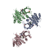
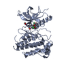
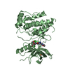
Mass: 551.383 Da / Num. of mol.: 3 / Source method: obtained synthetically / Formula: C22H24Cl2N8O5 / Feature type: SUBJECT OF INVESTIGATION
#3: Chemical ChemComp-SO4 / SULFATE ION / Sulfate


Mass: 96.063 Da / Num. of mol.: 2 / Source method: obtained synthetically / Formula: SO4
#4: Water ChemComp-HOH / water / Water


Mass: 18.015 Da / Num. of mol.: 4 / Source method: isolated from a natural source / Formula: H2O

-
Experimental details

-
Experiment

ExperimentMethod: X-RAY DIFFRACTION / Number of used crystals: 1

-
Sample preparation

CrystalDensity Matthews: 3.28 Å3/Da / Density % sol: 62.54 %
Crystal growTemperature: 277 K / Method: vapor diffusion, hanging drop / pH: 6.5
Details: 1:1 mix of protein + reservoir Protein at ~15 mg/mL formulated in: 20mM Tris pH 8, 20mM NaCl, 2mM TCEP Reservoir: 14-18% PEG 10K 0.3 M (NH4)2SO4 0.1 M MES pH 6.5 5% ethylene glycol

-
Data collection

DiffractionMean temperature: 120 K / Serial crystal experiment: N
Diffraction sourceSource: SYNCHROTRON / Site: APS / Beamline: 22-ID / Wavelength: 1 Å
DetectorType: RAYONIX MX300-HS / Detector: CCD / Date: Aug 14, 2014
RadiationProtocol: SINGLE WAVELENGTH / Monochromatic (M) / Laue (L): M / Scattering type: x-ray
Radiation wavelengthWavelength: 1 Å / Relative weight: 1
ReflectionResolution: 2.9→50 Å / Num. obs: 28812 / % possible obs: 94.2 % / Redundancy: 3.5 % / Rmerge(I) obs: 0.062 / Net I/σ(I): 19.3
Reflection shellResolution: 2.9→3 Å / Rmerge(I) obs: 0.29 / Mean I/σ(I) obs: 2.7 / Num. unique obs: 1973

-
Processing

Software
NameVersionClassification
REFMAC5.7.0029refinement
HKL-2000data reduction
SCALEPACKdata scaling
MOLREPphasing
RefinementMethod to determine structure: MOLECULAR REPLACEMENT
Starting model: 3RHX
Resolution: 2.9→48.28 Å / Cor.coef. Fo:Fc: 0.936 / Cor.coef. Fo:Fc free: 0.884 / SU B: 17.778 / SU ML: 0.341 / Cross valid method: THROUGHOUT / ESU R Free: 0.457 / Details: HYDROGENS HAVE BEEN ADDED IN THE RIDING POSITIONS
RfactorNum. reflection% reflectionSelection details
Rfree0.30265 1445 5 %RANDOM
Rwork0.22561 ---
obs0.22951 27367 94.05 %-
Solvent computationIon probe radii: 0.8 Å / Shrinkage radii: 0.8 Å / VDW probe radii: 1.2 Å
Displacement parametersBiso mean: 108.867 Å2
Baniso -1Baniso -2Baniso -3
1--0.02 Å20 Å20.13 Å2
2--0.23 Å20 Å2
3----0.2 Å2
Refinement stepCycle: 1 / Resolution: 2.9→48.28 Å
ProteinNucleic acidLigandSolventTotal
Num. atoms6788 0 121 4 6913
Refine LS restraints
Refine-IDTypeDev idealDev ideal targetNumber
X-RAY DIFFRACTIONr_bond_refined_d0.0110.027047
X-RAY DIFFRACTIONr_bond_other_d0.0020.026788
X-RAY DIFFRACTIONr_angle_refined_deg1.5162.0029528
X-RAY DIFFRACTIONr_angle_other_deg0.8843.00415654
X-RAY DIFFRACTIONr_dihedral_angle_1_deg6.8365850
X-RAY DIFFRACTIONr_dihedral_angle_2_deg38.68524.338302
X-RAY DIFFRACTIONr_dihedral_angle_3_deg17.983151267
X-RAY DIFFRACTIONr_dihedral_angle_4_deg16.2791545
X-RAY DIFFRACTIONr_chiral_restr0.0740.21041
X-RAY DIFFRACTIONr_gen_planes_refined0.0060.0217781
X-RAY DIFFRACTIONr_gen_planes_other0.0010.021481
X-RAY DIFFRACTIONr_nbd_refined
X-RAY DIFFRACTIONr_nbd_other
X-RAY DIFFRACTIONr_nbtor_refined
X-RAY DIFFRACTIONr_nbtor_other
X-RAY DIFFRACTIONr_xyhbond_nbd_refined
X-RAY DIFFRACTIONr_xyhbond_nbd_other
X-RAY DIFFRACTIONr_metal_ion_refined
X-RAY DIFFRACTIONr_metal_ion_other
X-RAY DIFFRACTIONr_symmetry_vdw_refined
X-RAY DIFFRACTIONr_symmetry_vdw_other
X-RAY DIFFRACTIONr_symmetry_hbond_refined
X-RAY DIFFRACTIONr_symmetry_hbond_other
X-RAY DIFFRACTIONr_symmetry_metal_ion_refined
X-RAY DIFFRACTIONr_symmetry_metal_ion_other
X-RAY DIFFRACTIONr_mcbond_it
X-RAY DIFFRACTIONr_mcbond_other
X-RAY DIFFRACTIONr_mcangle_it
X-RAY DIFFRACTIONr_mcangle_other
X-RAY DIFFRACTIONr_scbond_it
X-RAY DIFFRACTIONr_scbond_other
X-RAY DIFFRACTIONr_scangle_it
X-RAY DIFFRACTIONr_scangle_other
X-RAY DIFFRACTIONr_long_range_B_refined
X-RAY DIFFRACTIONr_long_range_B_other
X-RAY DIFFRACTIONr_rigid_bond_restr
X-RAY DIFFRACTIONr_sphericity_free
X-RAY DIFFRACTIONr_sphericity_bonded
LS refinement shellResolution: 2.896→2.971 Å / Total num. of bins used: 20
RfactorNum. reflection% reflection
Rfree0.424 74 -
Rwork0.333 1299 -
obs--60.97 %

+
About Yorodumi

-
News

-
Feb 9, 2022. New format data for meta-information of EMDB entries

New format data for meta-information of EMDB entries

  • Version 3 of the EMDB header file is now the official format.
  • The previous official version 1.9 will be removed from the archive.

Related info.:EMDB header

External links:wwPDB to switch to version 3 of the EMDB data model

-
Aug 12, 2020. Covid-19 info

Covid-19 info

URL: https://pdbj.org/emnavi/covid19.php

New page: Covid-19 featured information page in EM Navigator.

Related info.:Covid-19 info / Mar 5, 2020. Novel coronavirus structure data

+
Mar 5, 2020. Novel coronavirus structure data

Novel coronavirus structure data

Related info.:Yorodumi Speices / Aug 12, 2020. Covid-19 info

External links:COVID-19 featured content - PDBj / Molecule of the Month (242):Coronavirus Proteases

+
Jan 31, 2019. EMDB accession codes are about to change! (news from PDBe EMDB page)

EMDB accession codes are about to change! (news from PDBe EMDB page)

  • The allocation of 4 digits for EMDB accession codes will soon come to an end. Whilst these codes will remain in use, new EMDB accession codes will include an additional digit and will expand incrementally as the available range of codes is exhausted. The current 4-digit format prefixed with “EMD-” (i.e. EMD-XXXX) will advance to a 5-digit format (i.e. EMD-XXXXX), and so on. It is currently estimated that the 4-digit codes will be depleted around Spring 2019, at which point the 5-digit format will come into force.
  • The EM Navigator/Yorodumi systems omit the EMD- prefix.

Related info.:Q: What is EMD? / ID/Accession-code notation in Yorodumi/EM Navigator

External links:EMDB Accession Codes are Changing Soon! / Contact to PDBj

+
Jul 12, 2017. Major update of PDB

Major update of PDB

  • wwPDB released updated PDB data conforming to the new PDBx/mmCIF dictionary.
  • This is a major update changing the version number from 4 to 5, and with Remediation, in which all the entries are updated.
  • In this update, many items about electron microscopy experimental information are reorganized (e.g. em_software).
  • Now, EM Navigator and Yorodumi are based on the updated data.

External links:wwPDB Remediation / Enriched Model Files Conforming to OneDep Data Standards Now Available in the PDB FTP Archive

-
Yorodumi

Thousand views of thousand structures

  • Yorodumi is a browser for structure data from EMDB, PDB, SASBDB, etc.
  • This page is also the successor to EM Navigator detail page, and also detail information page/front-end page for Omokage search.
  • The word "yorodu" (or yorozu) is an old Japanese word meaning "ten thousand". "mi" (miru) is to see.

Related info.:EMDB / PDB / SASBDB / Comparison of 3 databanks / Yorodumi Search / Aug 31, 2016. New EM Navigator & Yorodumi / Yorodumi Papers / Jmol/JSmol / Function and homology information / Changes in new EM Navigator and Yorodumi

Read more