 Movie
Movie Controller

[English] 日本語
 Yorodumi
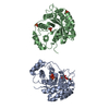
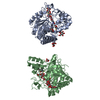
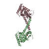
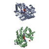
- PDB-6oov: Crystal structure of HMCES SRAP domain in complex with palindromi... -
+ Open data

- Basic information
| Entry | Database: PDB / ID: 6oov | ||||||
|---|---|---|---|---|---|---|---|
| Title | Crystal structure of HMCES SRAP domain in complex with palindromic 3' overhang DNA | ||||||
 Components Components |
| ||||||
 Keywords Keywords | DNA BINDING PROTEIN/DNA / SRAP Domain /  HMCES / Structural Genomics / Structural Genomics Consortium / SGC / DNA binding protein / DNA-protein complex / DNA-damage protein / 3' overhang / DNA BINDING PROTEIN-DNA complex HMCES / Structural Genomics / Structural Genomics Consortium / SGC / DNA binding protein / DNA-protein complex / DNA-damage protein / 3' overhang / DNA BINDING PROTEIN-DNA complex | ||||||
| Function / homology |  Function and homology information Function and homology information: / positive regulation of isotype switching / double-strand break repair via alternative nonhomologous end joining / Hydrolases; Acting on peptide bonds (peptidases) / replication fork / single-stranded DNA binding / peptidase activity / DNA damage response / proteolysisSimilarity search - Function | ||||||
| Biological species |  Homo sapiens (human) Homo sapiens (human)synthetic construct (others) | ||||||
| Method | X-RAY DIFFRACTION / SYNCHROTRON / FOURIER SYNTHESIS / Resolution: 2.2 Å | ||||||
 Authors Authors | Halabelian, L. / Zeng, H. / Li, Y. / Bountra, C. / Edwards, A.M. / Arrowsmith, C.H. / Structural Genomics Consortium (SGC) | ||||||
 Citation Citation | Journal: To Be Published Title: Crystal structure of HMCES SRAP domain in complex with palindromic 3' overhang DNA Authors: Halabelian, L. / Zeng, H. / Li, Y. / Bountra, C. / Edwards, A.M. / Arrowsmith, C.H. / Structural Genomics Consortium (SGC) | ||||||
| History |
|
- Structure visualization
| Structure viewer | Molecule: MolmilJmol/JSmol |
|---|
- Downloads & links
-Download
| PDBx/mmCIF format | 6oov.cif.gz | 243.3 KB | Display | PDBx/mmCIF format |
|---|---|---|---|---|
| PDB format | pdb6oov.ent.gz | 190.3 KB | Display | PDB format |
| PDBx/mmJSON format | 6oov.json.gz | Tree view | PDBx/mmJSON format | |
| Others |  Other downloads Other downloads |
-Validation report
| Arichive directory | https://data.pdbj.org/pub/pdb/validation_reports/oo/6oovftp://data.pdbj.org/pub/pdb/validation_reports/oo/6oov | HTTPS FTP |
|---|
-Related structure data
| Related structure data | 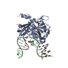 6oebS S: Starting model for refinement |
|---|---|
| Similar structure data |


-Links
 PDBj
PDBj


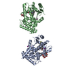
- Assembly
| Deposited unit | 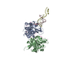
| ||||||||||||||||||
|---|---|---|---|---|---|---|---|---|---|---|---|---|---|---|---|---|---|---|---|
| 1 | 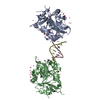
| ||||||||||||||||||
| Unit cell |
| ||||||||||||||||||
| Noncrystallographic symmetry (NCS) | NCS domain:
NCS domain segments: Component-ID: 0 / Ens-ID: 1 / Beg auth comp-ID: CYS / Beg label comp-ID: CYS / End auth comp-ID: VAL / End label comp-ID: VAL / Refine code: 0 / Auth seq-ID: 2 - 270 / Label seq-ID: 1 - 269
|
-Components
| #1: Protein | Mass: 31671.617 Da / Num. of mol.: 2 Source method: isolated from a genetically manipulated source Source: (gene. exp.) Homo sapiens (human) / Gene: HMCES, C3orf37, DC12, SRAPD1 / Production host:  Escherichia coli (E. coli) Escherichia coli (E. coli)References: UniProt: Q96FZ2, Hydrolases; Acting on peptide bonds (peptidases)#2: DNA chain | Mass: 3947.581 Da / Num. of mol.: 2 / Source method: obtained synthetically / Source: (synth.) synthetic construct (others) #3: Chemical | ChemComp-UNX /   Num. of mol.: 47 / Source method: obtained synthetically Num. of mol.: 47 / Source method: obtained synthetically#4: Water | ChemComp-HOH / | Water Mass: 18.015 Da / Num. of mol.: 82 / Source method: isolated from a natural source / Formula: H2O Mass: 18.015 Da / Num. of mol.: 82 / Source method: isolated from a natural source / Formula: H2O |
|---|
-Experimental details
-Experiment
| Experiment | Method: X-RAY DIFFRACTION / Number of used crystals: 1 |
|---|
- Sample preparation
| Crystal | Density Matthews: 2.94 Å3/Da / Density % sol: 58.22 % |
|---|---|
| Crystal grow | Temperature: 293 K / Method: vapor diffusion, sitting drop / pH: 7.5 / Details: 11% PEG 8K, 0.1M CaCl2, 0.1M Hepes |
-Data collection
| Diffraction | Mean temperature: 100 K / Serial crystal experiment: N | |||||||||||||||||||||||||||
|---|---|---|---|---|---|---|---|---|---|---|---|---|---|---|---|---|---|---|---|---|---|---|---|---|---|---|---|---|
| Diffraction source | Source: SYNCHROTRON / Site: ALS  / Beamline: 5.0.1 / Wavelength: 0.97741 Å / Beamline: 5.0.1 / Wavelength: 0.97741 Å | |||||||||||||||||||||||||||
| Detector | Type: DECTRIS PILATUS3 6M / Detector: PIXEL / Date: Sep 18, 2018 | |||||||||||||||||||||||||||
| Radiation | Protocol: SINGLE WAVELENGTH / Monochromatic (M) / Laue (L): M / Scattering type: x-ray | |||||||||||||||||||||||||||
| Radiation wavelength | Wavelength: 0.97741 Å / Relative weight: 1 | |||||||||||||||||||||||||||
| Reflection | Resolution: 2.2→47.84 Å / Num. obs: 41672 / % possible obs: 99.7 % / Redundancy: 3.4 % / CC1/2: 0.999 / Rmerge(I) obs: 0.056 / Rpim(I) all: 0.035 / Rrim(I) all: 0.066 / Net I/σ(I): 13.8 / Num. measured all: 142073 / Scaling rejects: 5 | |||||||||||||||||||||||||||
| Reflection shell | Diffraction-ID: 1 / Redundancy: 3.3 %
|
- Processing
| Software |
| |||||||||||||||||||||||||||||||||||||||||||||||||||||||||||||||||||||||||||||||||||||||||||||||||||||||||||||||||||||||||||||
|---|---|---|---|---|---|---|---|---|---|---|---|---|---|---|---|---|---|---|---|---|---|---|---|---|---|---|---|---|---|---|---|---|---|---|---|---|---|---|---|---|---|---|---|---|---|---|---|---|---|---|---|---|---|---|---|---|---|---|---|---|---|---|---|---|---|---|---|---|---|---|---|---|---|---|---|---|---|---|---|---|---|---|---|---|---|---|---|---|---|---|---|---|---|---|---|---|---|---|---|---|---|---|---|---|---|---|---|---|---|---|---|---|---|---|---|---|---|---|---|---|---|---|---|---|---|---|
| Refinement | Method to determine structure: FOURIER SYNTHESIS Starting model: 6OEB Resolution: 2.2→47.84 Å / Cor.coef. Fo:Fc: 0.961 / Cor.coef. Fo:Fc free: 0.951 / SU B: 14.2 / SU ML: 0.162 / SU R Cruickshank DPI: 0.1992 / Cross valid method: THROUGHOUT / σ(F): 0 / ESU R: 0.199 / ESU R Free: 0.166 Details: HYDROGENS HAVE BEEN ADDED IN THE RIDING POSITIONS U VALUES : WITH TLS ADDED
| |||||||||||||||||||||||||||||||||||||||||||||||||||||||||||||||||||||||||||||||||||||||||||||||||||||||||||||||||||||||||||||
| Solvent computation | Ion probe radii: 0.8 Å / Shrinkage radii: 0.8 Å / VDW probe radii: 1.2 Å | |||||||||||||||||||||||||||||||||||||||||||||||||||||||||||||||||||||||||||||||||||||||||||||||||||||||||||||||||||||||||||||
| Displacement parameters | Biso max: 156.62 Å2 / Biso mean: 55.49 Å2 / Biso min: 15.57 Å2
| |||||||||||||||||||||||||||||||||||||||||||||||||||||||||||||||||||||||||||||||||||||||||||||||||||||||||||||||||||||||||||||
| Refinement step | Cycle: final / Resolution: 2.2→47.84 Å
| |||||||||||||||||||||||||||||||||||||||||||||||||||||||||||||||||||||||||||||||||||||||||||||||||||||||||||||||||||||||||||||
| Refine LS restraints |
| |||||||||||||||||||||||||||||||||||||||||||||||||||||||||||||||||||||||||||||||||||||||||||||||||||||||||||||||||||||||||||||
| Refine LS restraints NCS | Ens-ID: 1 / Number: 7881 / Refine-ID: X-RAY DIFFRACTION / Type: interatomic distance / Rms dev position: 0.07 Å / Weight position: 0.05
| |||||||||||||||||||||||||||||||||||||||||||||||||||||||||||||||||||||||||||||||||||||||||||||||||||||||||||||||||||||||||||||
| LS refinement shell | Resolution: 2.2→2.257 Å / Rfactor Rfree error: 0 / Total num. of bins used: 20
| |||||||||||||||||||||||||||||||||||||||||||||||||||||||||||||||||||||||||||||||||||||||||||||||||||||||||||||||||||||||||||||
| Refinement TLS params. | Method: refined / Refine-ID: X-RAY DIFFRACTION
| |||||||||||||||||||||||||||||||||||||||||||||||||||||||||||||||||||||||||||||||||||||||||||||||||||||||||||||||||||||||||||||
| Refinement TLS group |
|
