 Movie
Movie Controller

[English] 日本語
 Yorodumi
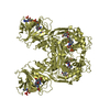
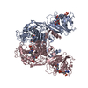
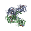
- PDB-6ojs: Crystal structure of TS-DHFR from Cryptosporidium hominis in comp... -
+ Open data

- Basic information
| Entry | Database: PDB / ID: 6ojs | |||||||||
|---|---|---|---|---|---|---|---|---|---|---|
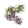
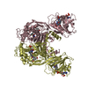
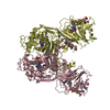
| Title | Crystal structure of TS-DHFR from Cryptosporidium hominis in complex with NADPH, FdUMP, MTX and 2-amino-4-oxo-4,7-dihydro-pyrrolo[2,3-d]pyrimidine-methyl-phenyl-D-glutamic acid | |||||||||
 Components Components | Bifunctional dihydrofolate reductase-thymidylate synthase | |||||||||
 Keywords Keywords | TRANSFERASE/TRANSFERASE INHIBITOR / Inhibitor / TS / TS-DHFR /  TRANSFERASE / TRANSFERASE-TRANSFERASE INHIBITOR complex TRANSFERASE / TRANSFERASE-TRANSFERASE INHIBITOR complex | |||||||||
| Function / homology |  Function and homology informationthymidylate synthase activity / dTMP biosynthetic process / dihydrofolate reductase activity / tetrahydrofolate biosynthetic process / one-carbon metabolic process / methylation Function and homology informationthymidylate synthase activity / dTMP biosynthetic process / dihydrofolate reductase activity / tetrahydrofolate biosynthetic process / one-carbon metabolic process / methylationSimilarity search - Function | |||||||||
| Biological species |  Cryptosporidium hominis (eukaryote) Cryptosporidium hominis (eukaryote) | |||||||||
| Method | X-RAY DIFFRACTION / SYNCHROTRON / MOLECULAR REPLACEMENT / Resolution: 3.214 Å | |||||||||
 Authors Authors | Czyzyk, D.J. / Anderson, K.S. / Jorgensen, W.L. / Valhondo, M. | |||||||||
| Funding support |  United States, 2items United States, 2items
| |||||||||
 Citation Citation | Journal: Febs Lett. / Year: 2019 Title: Understanding the structural basis of species selective, stereospecific inhibition for Cryptosporidium and human thymidylate synthase. Authors: Czyzyk, D.J. / Valhondo, M. / Jorgensen, W.L. / Anderson, K.S. | |||||||||
| History |
|
- Structure visualization
| Structure viewer | Molecule: MolmilJmol/JSmol |
|---|
- Downloads & links
-Download
| PDBx/mmCIF format | 6ojs.cif.gz | 520.3 KB | Display | PDBx/mmCIF format |
|---|---|---|---|---|
| PDB format | pdb6ojs.ent.gz | 424.7 KB | Display | PDB format |
| PDBx/mmJSON format | 6ojs.json.gz | Tree view | PDBx/mmJSON format | |
| Others |  Other downloads Other downloads |
-Validation report
| Arichive directory | https://data.pdbj.org/pub/pdb/validation_reports/oj/6ojsftp://data.pdbj.org/pub/pdb/validation_reports/oj/6ojs | HTTPS FTP |
|---|
-Related structure data
| Related structure data | 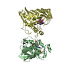 6ojuC  6ojvC  4q0dS S: Starting model for refinement C: citing same article ( |
|---|---|
| Similar structure data |


-Links
 PDBj
PDBj

- Assembly
| Deposited unit | 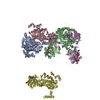
| ||||||||
|---|---|---|---|---|---|---|---|---|---|
| 1 | 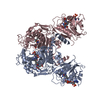
| ||||||||
| 2 | 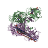
| ||||||||
| 3 | 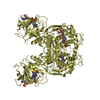
| ||||||||
| Unit cell |
|
-Components
| #1: Protein | Mass: 60262.520 Da / Num. of mol.: 5 Source method: isolated from a genetically manipulated source Source: (gene. exp.) Cryptosporidium hominis (eukaryote) / Gene: CHUDEA4_4460 / Production host:  Escherichia coli BL21(DE3) (bacteria) / Strain (production host): BL21(DE3) / Variant (production host): PA-414 / References: UniProt: A0A0S4TER9 Escherichia coli BL21(DE3) (bacteria) / Strain (production host): BL21(DE3) / Variant (production host): PA-414 / References: UniProt: A0A0S4TER9#2: Chemical | ChemComp-NDP / Nicotinamide adenine dinucleotide phosphate  Mass: 745.421 Da / Num. of mol.: 5 / Source method: obtained synthetically / Formula: C21H30N7O17P3 Mass: 745.421 Da / Num. of mol.: 5 / Source method: obtained synthetically / Formula: C21H30N7O17P3#3: Chemical | ChemComp-UFP / Fluorodeoxyuridylate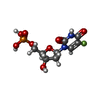  Type: DNA linking / Mass: 326.172 Da / Num. of mol.: 5 / Source method: obtained synthetically / Formula: C9H12FN2O8P / Comment: inhibitor*YM Type: DNA linking / Mass: 326.172 Da / Num. of mol.: 5 / Source method: obtained synthetically / Formula: C9H12FN2O8P / Comment: inhibitor*YM#4: Chemical | ChemComp-D96 /   Mass: 413.384 Da / Num. of mol.: 5 / Source method: obtained synthetically / Formula: C19H19N5O6 Mass: 413.384 Da / Num. of mol.: 5 / Source method: obtained synthetically / Formula: C19H19N5O6#5: Chemical | ChemComp-MTX / Methotrexate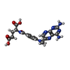  Mass: 454.439 Da / Num. of mol.: 5 / Source method: obtained synthetically / Formula: C20H22N8O5 / Comment: chemotherapy*YM Mass: 454.439 Da / Num. of mol.: 5 / Source method: obtained synthetically / Formula: C20H22N8O5 / Comment: chemotherapy*YM |
|---|
-Experimental details
-Experiment
| Experiment | Method: X-RAY DIFFRACTION / Number of used crystals: 1 |
|---|
- Sample preparation
| Crystal | Density Matthews: 4.63 Å3/Da / Density % sol: 73.5 % |
|---|---|
| Crystal grow | Temperature: 295.15 K / Method: vapor diffusion, hanging drop / pH: 8 Details: Well solution 22% PEG 6000, 0.2 M ammonium sulfate, 0.06 M lithium sulfate, 0.1 M Tris Drop ratio 2:1 Enzyme mix/well solution |
-Data collection
| Diffraction | Mean temperature: 100 K / Serial crystal experiment: N |
|---|---|
| Diffraction source | Source: SYNCHROTRON / Site: APS / Beamline: 24-ID-E / Wavelength: 0.97915 Å |
| Detector | Type: DECTRIS EIGER X 16M / Detector: PIXEL / Date: Jul 6, 2018 |
| Radiation | Protocol: SINGLE WAVELENGTH / Monochromatic (M) / Laue (L): M / Scattering type: x-ray |
| Radiation wavelength | Wavelength: 0.97915 Å / Relative weight: 1 |
| Reflection | Resolution: 3.21→51 Å / Num. obs: 85851 / % possible obs: 98.2 % / Redundancy: 6.9 % / CC1/2: 0.983 / Rsym value: 0.317 / Net I/σ(I): 4.98 |
| Reflection shell | Resolution: 3.21→3.41 Å / Redundancy: 6.7 % / Mean I/σ(I) obs: 1.06 / Num. unique obs: 12774 / CC1/2: 0.616 / Rsym value: 1.734 / % possible all: 91.1 |
- Processing
| Software |
| |||||||||||||||||||||||||||||||||||||||||||||||||||||||||||||||||||||||||||||||||||||||||||||||||||||||||||||||||||||||||||||||||||||||||||||||||||||||||||||||||||||||||||||||||||||||||||||||||||||||||||||||||||||||||
|---|---|---|---|---|---|---|---|---|---|---|---|---|---|---|---|---|---|---|---|---|---|---|---|---|---|---|---|---|---|---|---|---|---|---|---|---|---|---|---|---|---|---|---|---|---|---|---|---|---|---|---|---|---|---|---|---|---|---|---|---|---|---|---|---|---|---|---|---|---|---|---|---|---|---|---|---|---|---|---|---|---|---|---|---|---|---|---|---|---|---|---|---|---|---|---|---|---|---|---|---|---|---|---|---|---|---|---|---|---|---|---|---|---|---|---|---|---|---|---|---|---|---|---|---|---|---|---|---|---|---|---|---|---|---|---|---|---|---|---|---|---|---|---|---|---|---|---|---|---|---|---|---|---|---|---|---|---|---|---|---|---|---|---|---|---|---|---|---|---|---|---|---|---|---|---|---|---|---|---|---|---|---|---|---|---|---|---|---|---|---|---|---|---|---|---|---|---|---|---|---|---|---|---|---|---|---|---|---|---|---|---|---|---|---|---|---|---|---|
| Refinement | Method to determine structure: MOLECULAR REPLACEMENT Starting model: 4Q0D Resolution: 3.214→50.121 Å / SU ML: 0.45 / Cross valid method: FREE R-VALUE / σ(F): 1.33 / Phase error: 27.19
| |||||||||||||||||||||||||||||||||||||||||||||||||||||||||||||||||||||||||||||||||||||||||||||||||||||||||||||||||||||||||||||||||||||||||||||||||||||||||||||||||||||||||||||||||||||||||||||||||||||||||||||||||||||||||
| Solvent computation | Shrinkage radii: 0.9 Å / VDW probe radii: 1.11 Å | |||||||||||||||||||||||||||||||||||||||||||||||||||||||||||||||||||||||||||||||||||||||||||||||||||||||||||||||||||||||||||||||||||||||||||||||||||||||||||||||||||||||||||||||||||||||||||||||||||||||||||||||||||||||||
| Refinement step | Cycle: LAST / Resolution: 3.214→50.121 Å
| |||||||||||||||||||||||||||||||||||||||||||||||||||||||||||||||||||||||||||||||||||||||||||||||||||||||||||||||||||||||||||||||||||||||||||||||||||||||||||||||||||||||||||||||||||||||||||||||||||||||||||||||||||||||||
| Refine LS restraints |
| |||||||||||||||||||||||||||||||||||||||||||||||||||||||||||||||||||||||||||||||||||||||||||||||||||||||||||||||||||||||||||||||||||||||||||||||||||||||||||||||||||||||||||||||||||||||||||||||||||||||||||||||||||||||||
| LS refinement shell |
|
