Movie
Movie Controller


+ Open data

- Basic information
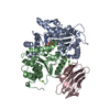
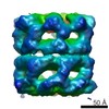
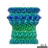
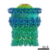
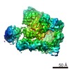
| Entry | Database: PDB / ID: 6oif | |||||||||
|---|---|---|---|---|---|---|---|---|---|---|
| Title | Cryo-EM structure of human TorsinA filament | |||||||||
 Components Components | Torsin-1A | |||||||||
 Keywords Keywords |  HYDROLASE / AAA+ ATPase / nucleotide binding / nuclear envelope / endoplasmic reticulum / membrane HYDROLASE / AAA+ ATPase / nucleotide binding / nuclear envelope / endoplasmic reticulum / membrane | |||||||||
| Function / homology |  Function and homology information Function and homology informationsynaptic vesicle membrane organization / nuclear membrane organization / organelle organization / regulation of dopamine uptake involved in synaptic transmission / nuclear envelope organization / intermediate filament cytoskeleton organization / protein deneddylation / regulation of protein localization to cell surface / positive regulation of synaptic vesicle endocytosis / misfolded protein binding ...synaptic vesicle membrane organization / nuclear membrane organization / organelle organization / regulation of dopamine uptake involved in synaptic transmission / nuclear envelope organization / intermediate filament cytoskeleton organization / protein deneddylation / regulation of protein localization to cell surface / positive regulation of synaptic vesicle endocytosis / misfolded protein binding / : / wound healing, spreading of cells / synaptic vesicle transport / chaperone cofactor-dependent protein refolding / kinesin binding / protein localization to nucleus / chaperone-mediated protein folding / cytoskeletal protein binding / secretory granule / ATP-dependent protein folding chaperone / Hydrolases; Acting on acid anhydrides; Acting on acid anhydrides to facilitate cellular and subcellular movement / cytoplasmic vesicle membrane / neuron projection development / unfolded protein binding / synaptic vesicle / Cargo recognition for clathrin-mediated endocytosis / nuclear envelope / growth cone / nuclear membrane / response to oxidative stress / cytoskeleton / cell adhesion / endoplasmic reticulum lumen / intracellular membrane-bounded organelle / endoplasmic reticulum membrane / endoplasmic reticulum / ATP hydrolysis activity / extracellular exosome / ATP binding / membrane / identical protein binding / cytosolSimilarity search - Function | |||||||||
| Biological species |  Homo sapiens (human) Homo sapiens (human) | |||||||||
| Method | ELECTRON MICROSCOPY / helical reconstruction / cryo EM / Resolution: 4.4 Å | |||||||||
 Authors Authors | Zheng, W. / Demircioglu, F.E. / Schwartz, T.U. / Egelman, E.H. | |||||||||
| Funding support |  United States, 2items United States, 2items
| |||||||||
 Citation Citation | Journal: Nat Commun / Year: 2019 Title: The AAA + ATPase TorsinA polymerizes into hollow helical tubes with 8.5 subunits per turn. Authors: F Esra Demircioglu / Weili Zheng / Alexander J McQuown / Nolan K Maier / Nicki Watson / Iain M Cheeseman / Vladimir Denic / Edward H Egelman / Thomas U Schwartz / Abstract: TorsinA is an ER-resident AAA + ATPase, whose deletion of glutamate E303 results in the genetic neuromuscular disease primary dystonia. TorsinA is an unusual AAA + ATPase that needs an ...TorsinA is an ER-resident AAA + ATPase, whose deletion of glutamate E303 results in the genetic neuromuscular disease primary dystonia. TorsinA is an unusual AAA + ATPase that needs an external activator. Also, it likely does not thread a peptide substrate through a narrow central channel, in contrast to its closest structural homologs. Here, we examined the oligomerization of TorsinA to get closer to a molecular understanding of its still enigmatic function. We observe TorsinA to form helical filaments, which we analyzed by cryo-electron microscopy using helical reconstruction. The 4.4 Å structure reveals long hollow tubes with a helical periodicity of 8.5 subunits per turn, and an inner channel of ~ 4 nm diameter. We further show that the protein is able to induce tubulation of membranes in vitro, an observation that may reflect an entirely new characteristic of AAA + ATPases. We discuss the implications of these observations for TorsinA function. | |||||||||
| History |
|
- Structure visualization
| Movie |
Movie viewer |
|---|---|
| Structure viewer | Molecule: MolmilJmol/JSmol |
- Downloads & links
-Download
| PDBx/mmCIF format | 6oif.cif.gz | 2.2 MB | Display | PDBx/mmCIF format |
|---|---|---|---|---|
| PDB format | pdb6oif.ent.gz | Display | PDB format | |
| PDBx/mmJSON format | 6oif.json.gz | Tree view | PDBx/mmJSON format | |
| Others |  Other downloads Other downloads |
-Validation report
| Arichive directory | https://data.pdbj.org/pub/pdb/validation_reports/oi/6oifftp://data.pdbj.org/pub/pdb/validation_reports/oi/6oif | HTTPS FTP |
|---|
-Related structure data
| Related structure data | 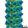 20076MC M: map data used to model this data C: citing same article ( |
|---|---|
| Similar structure data |
-Links
 PDBj
PDBj- Assembly
| Deposited unit | 
|
|---|---|
| 1 |
|
| Symmetry | Helical symmetry: (Circular symmetry: 1 / Dyad axis: no / N subunits divisor: 1 / Num. of operations: 25 / Rise per n subunits: 5.53 Å / Rotation per n subunits: 42.51 °) |
-Components
| #1: Protein | Mass: 32590.248 Da / Num. of mol.: 25 Source method: isolated from a genetically manipulated source Source: (gene. exp.) Homo sapiens (human) / Gene: TOR1A, DQ2, DYT1, TA, TORA / Production host:  Escherichia coli (E. coli) Escherichia coli (E. coli)References: UniProt: O14656, Hydrolases; Acting on acid anhydrides; Acting on acid anhydrides to facilitate cellular and subcellular movement#2: Chemical | ChemComp-ATP / Adenosine triphosphate  Mass: 507.181 Da / Num. of mol.: 25 / Source method: obtained synthetically / Formula: C10H16N5O13P3 / Comment: ATP, energy-carrying molecule*YM Mass: 507.181 Da / Num. of mol.: 25 / Source method: obtained synthetically / Formula: C10H16N5O13P3 / Comment: ATP, energy-carrying molecule*YM |
|---|
-Experimental details
-Experiment
| Experiment | Method: ELECTRON MICROSCOPY |
|---|---|
| EM experiment | Aggregation state: FILAMENT / 3D reconstruction method: helical reconstruction |
- Sample preparation
| Component | Name: TorsinA / Type: COMPLEX / Entity ID: #1 / Source: RECOMBINANT |
|---|---|
| Source (natural) | Organism: Homo sapiens (human) |
| Source (recombinant) | Organism: Escherichia coli (E. coli) |
| Buffer solution | pH: 8 |
| Specimen | Embedding applied: NO / Shadowing applied: NO / Staining applied: NO / Vitrification applied: YES |
| Specimen support | Details: unspecified |
| Vitrification | Cryogen name: ETHANE |
- Electron microscopy imaging
| Experimental equipment |  Model: Talos Arctica / Image courtesy: FEI Company |
|---|---|
| Microscopy | Model: FEI TALOS ARCTICA |
| Electron gun | Electron source: FIELD EMISSION GUN / Accelerating voltage: 200 kV / Illumination mode: FLOOD BEAM |
| Electron lens | Mode: BRIGHT FIELDBright-field microscopy |
| Image recording | Electron dose: 30 e/Å2 / Detector mode: COUNTING / Film or detector model: GATAN K2 SUMMIT (4k x 4k) |
- Processing
| EM software |
| ||||||||||||||||||||||||||||||||||||||||||||||||||
|---|---|---|---|---|---|---|---|---|---|---|---|---|---|---|---|---|---|---|---|---|---|---|---|---|---|---|---|---|---|---|---|---|---|---|---|---|---|---|---|---|---|---|---|---|---|---|---|---|---|---|---|
| CTF correction | Type: PHASE FLIPPING AND AMPLITUDE CORRECTION | ||||||||||||||||||||||||||||||||||||||||||||||||||
| Helical symmerty | Angular rotation/subunit: 42.51 ° / Axial rise/subunit: 5.53 Å / Axial symmetry: C1 | ||||||||||||||||||||||||||||||||||||||||||||||||||
| Particle selection | Num. of particles selected: 75909 | ||||||||||||||||||||||||||||||||||||||||||||||||||
| 3D reconstruction | Resolution: 4.4 Å / Resolution method: FSC 0.143 CUT-OFF / Num. of particles: 75909 / Symmetry type: HELICAL | ||||||||||||||||||||||||||||||||||||||||||||||||||
| Atomic model building | PDB-ID: 5J1S |