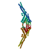
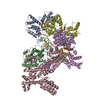
Entry Database : PDB / ID : 6o8cTitle Crystal structure of STING CTT in complex with TBK1 Serine/threonine-protein kinase TBK1 Stimulator of interferon genes protein Keywords / / / Function / homology Function Domain/homology Component
/ / / / / / / / / / / / / / / / / / / / / / / / / / / / / / / / / / / / / / / / / / / / / / / / / / / / / / / / / / / / / / / / / / / / / / / / / / / / / / / / / / / / / / / / / / / / / / / / / / / / / / / / / / / / / / / / / / / / / / / / / / / / / / / / / / / / Biological species Mus musculus (house mouse)Homo sapiens (human)Method / / / Resolution : 3.17 Å Authors Li, P. / Zhao, B. / Du, F. Funding support Organization Grant number Country Welch Foundation A-1931-20170325
Journal : Nature / Year : 2019Title : A conserved PLPLRT/SD motif of STING mediates the recruitment and activation of TBK1.Authors : Zhao, B. / Du, F. / Xu, P. / Shu, C. / Sankaran, B. / Bell, S.L. / Liu, M. / Lei, Y. / Gao, X. / Fu, X. / Zhu, F. / Liu, Y. / Laganowsky, A. / Zheng, X. / Ji, J.Y. / West, A.P. / Watson, R.O. / Li, P. History Deposition Mar 9, 2019 Deposition site / Processing site Revision 1.0 May 22, 2019 Provider / Type Revision 1.1 Jun 5, 2019 Group / Database references / Category / citation_authorItem / _citation.title / _citation_author.nameRevision 1.2 Jun 12, 2019 Group / Database references / Category Item / _citation.page_first / _citation.page_lastRevision 1.3 Oct 11, 2023 Group / Database references / Refinement descriptionCategory chem_comp_atom / chem_comp_bond ... chem_comp_atom / chem_comp_bond / database_2 / pdbx_initial_refinement_model Item / _database_2.pdbx_database_accession
Show all Show less
 Movie
Movie Controller


 Open data
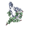
 Basic information
 Components
 Keywords
 IMMUNE SYSTEM /
  Function and homology information

 Authors
 United States, 1items
  Citation
 Structure visualization
 Downloads & links
 Other downloads


 PDBj
PDBj


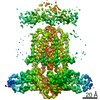
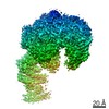
 Assembly


 Mass: 591.468 Da / Num. of mol.: 2 / Source method: obtained synthetically / Formula: C23H26IN7O2S
 Sample preparation
 Processing