| Entry | Database: PDB / ID: 6o54
|
|---|
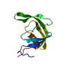
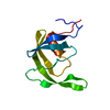
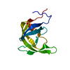
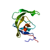
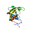
| Title | Crystal Structure of multi-drug resistant HIV-1 protease PR-S17 (D25N) |
|---|
 Components Components | HIV-1 protease |
|---|
 Keywords Keywords | HYDROLASE / Viral Protein / HIV PROTEASE |
|---|
| Function / homology |  Function and homology information Function and homology information
Retropepsin-like catalytic domain / Retropepsins / Retroviral aspartyl protease / Aspartyl protease, retroviral-type family profile. / Peptidase A2A, retrovirus, catalytic / Cathepsin D, subunit A; domain 1 / Acid Proteases / Aspartic peptidase, active site / Eukaryotic and viral aspartyl proteases active site. / Aspartic peptidase domain superfamily ...Retropepsin-like catalytic domain / Retropepsins / Retroviral aspartyl protease / Aspartyl protease, retroviral-type family profile. / Peptidase A2A, retrovirus, catalytic / Cathepsin D, subunit A; domain 1 / Acid Proteases / Aspartic peptidase, active site / Eukaryotic and viral aspartyl proteases active site. / Aspartic peptidase domain superfamily / Beta Barrel / Mainly BetaSimilarity search - Domain/homology |
|---|
| Biological species |   Human immunodeficiency virus 1 Human immunodeficiency virus 1 |
|---|
| Method | X-RAY DIFFRACTION / SYNCHROTRON / MOLECULAR REPLACEMENT / Resolution: 1.21 Å |
|---|
 Authors Authors | Wang, Y.-F. / Brothers, R. / Agniswamy, J. / Weber, I.T. |
|---|
| Funding support |  United States, 1items United States, 1items | Organization | Grant number | Country |
|---|
| National Institutes of Health/National Human Genome Research Institute (NIH/NHGRI) | GM062920 | United States |
|
|---|
 Citation Citation | Journal: Acs Omega / Year: 2019
Title: Highly Drug-Resistant HIV-1 Protease Mutant PRS17 Shows Enhanced Binding to Substrate Analogues.
Authors: Agniswamy, J. / Kneller, D.W. / Brothers, R. / Wang, Y.F. / Harrison, R.W. / Weber, I.T. |
|---|
| History | | Deposition | Mar 1, 2019 | Deposition site: RCSB / Processing site: RCSB |
|---|
| Revision 1.0 | Jun 19, 2019 | Provider: repository / Type: Initial release |
|---|
| Revision 1.1 | Jul 17, 2019 | Group: Data collection / Refinement description / Category: software
Item: _software.classification / _software.name / _software.version |
|---|
| Revision 1.2 | Dec 18, 2019 | Group: Author supporting evidence / Category: pdbx_audit_support / Item: _pdbx_audit_support.funding_organization |
|---|
| Revision 1.3 | Oct 11, 2023 | Group: Data collection / Database references / Refinement description
Category: chem_comp_atom / chem_comp_bond ...chem_comp_atom / chem_comp_bond / database_2 / pdbx_initial_refinement_model
Item: _database_2.pdbx_DOI / _database_2.pdbx_database_accession |
|---|
|
|---|
 Movie
Movie Controller

 Yorodumi
 Open data
 Basic information
 Structure visualization
 Downloads & links
 Other downloads


 PDBj
PDBj Assembly


 Mass: 35.453 Da / Num. of mol.: 4 / Source method: obtained synthetically / Formula: Cl
 Mass: 18.015 Da / Num. of mol.: 105 / Source method: isolated from a natural source / Formula: H2O
 Sample preparation
 Processing