 Movie
Movie Controller


+ Open data

- Basic information
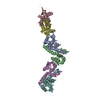
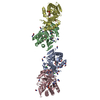
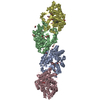
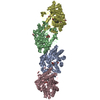
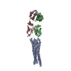
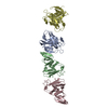
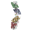
| Entry | Database: PDB / ID: 6o3n | |||||||||
|---|---|---|---|---|---|---|---|---|---|---|
| Title | Cross-alpha Amyloid-like Structure alphaAmA | |||||||||
 Components Components | Cross-alpha Amyloid-like Structure alphaAmA | |||||||||
 Keywords Keywords |  DE NOVO PROTEIN / alpha-amyloid / De Novo Design / coiled-coil / fibril DE NOVO PROTEIN / alpha-amyloid / De Novo Design / coiled-coil / fibril | |||||||||
| Biological species | synthetic (others) | |||||||||
| Method | X-RAY DIFFRACTION / SYNCHROTRON / MOLECULAR REPLACEMENT / Resolution: 3.7 Å | |||||||||
 Authors Authors | Zhang, S.-Q. / Liu, L. / DeGrado, W.F. | |||||||||
| Funding support |  United States, 2items United States, 2items
| |||||||||
 Citation Citation | Journal: Nat. Chem. Biol. / Year: 2018 Title: Designed peptides that assemble into cross-alpha amyloid-like structures Authors: Zhang, S.Q. / Huang, H. / Yang, J. / Kratochvil, H.T. / Lolicato, M. / Liu, Y. / Shu, X. / Liu, L. / DeGrado, W.F. | |||||||||
| History |
|
- Structure visualization
| Structure viewer | Molecule:  MolmilJmol/JSmol MolmilJmol/JSmol |
|---|
- Downloads & links
-Download
| PDBx/mmCIF format | 6o3n.cif.gz | 65.9 KB | Display | PDBx/mmCIF format |
|---|---|---|---|---|
| PDB format | pdb6o3n.ent.gz | 47.5 KB | Display | PDB format |
| PDBx/mmJSON format | 6o3n.json.gz | Tree view | PDBx/mmJSON format | |
| Others |  Other downloads Other downloads |
-Validation report
| Arichive directory | https://data.pdbj.org/pub/pdb/validation_reports/o3/6o3nftp://data.pdbj.org/pub/pdb/validation_reports/o3/6o3n | HTTPS FTP |
|---|
-Related structure data
| Related structure data |  6c4xC  6c4yC  6c4zSC  6c50C 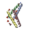 6c51C  6c52C 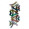 6d02C C: citing same article ( S: Starting model for refinement |
|---|---|
| Similar structure data |


-Links
 PDBj
PDBj
- Assembly
| Deposited unit | 
| ||||||||
|---|---|---|---|---|---|---|---|---|---|
| 1 | 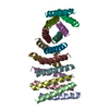
| ||||||||
| Unit cell |
|
-Components
| #1: Protein/peptide | Mass: 2932.569 Da / Num. of mol.: 16 / Source method: obtained synthetically / Source: (synth.) synthetic (others) |
|---|
-Experimental details
-Experiment
| Experiment | Method: X-RAY DIFFRACTION / Number of used crystals: 1 |
|---|
- Sample preparation
| Crystal | Density Matthews: 5.94 Å3/Da / Density % sol: 79.29 % |
|---|---|
| Crystal grow | Temperature: 298 K / Method: vapor diffusion, hanging drop / Details: 45% MPD, 0.25M NaH2PO4 |
-Data collection
| Diffraction | Mean temperature: 100 K / Serial crystal experiment: N | ||||||||||||||||||||
|---|---|---|---|---|---|---|---|---|---|---|---|---|---|---|---|---|---|---|---|---|---|
| Diffraction source | Source: SYNCHROTRON / Site: ALS / Beamline: 8.3.1 / Wavelength: 1.11583 Å | ||||||||||||||||||||
| Detector | Type: DECTRIS PILATUS3 S 6M / Detector: PIXEL / Date: Apr 12, 2017 | ||||||||||||||||||||
| Radiation | Protocol: SINGLE WAVELENGTH / Monochromatic (M) / Laue (L): M / Scattering type: x-ray | ||||||||||||||||||||
| Radiation wavelength | Wavelength: 1.11583 Å / Relative weight: 1 | ||||||||||||||||||||
| Reflection twin |
| ||||||||||||||||||||
| Reflection | Resolution: 3.7→116.5 Å / Num. obs: 12514 / % possible obs: 99.7 % / Redundancy: 25.6 % / CC1/2: 0.997 / Rrim(I) all: 0.098 / Net I/σ(I): 18.48 | ||||||||||||||||||||
| Reflection shell | Resolution: 3.7→3.93 Å / Redundancy: 25.5 % / Mean I/σ(I) obs: 1.59 / Num. unique obs: 1946 / CC1/2: 0.465 / Rrim(I) all: 2.11 / % possible all: 98 |
- Processing
| Software |
| ||||||||||||||||||||||||||||||||||||||||||||||||||||||||||||||||||||||||||||||||||||||||||||||||||||||||||||||||||||||||||||||||||||||||||||||||||||||||||||||||||||||||||||||||||||||
|---|---|---|---|---|---|---|---|---|---|---|---|---|---|---|---|---|---|---|---|---|---|---|---|---|---|---|---|---|---|---|---|---|---|---|---|---|---|---|---|---|---|---|---|---|---|---|---|---|---|---|---|---|---|---|---|---|---|---|---|---|---|---|---|---|---|---|---|---|---|---|---|---|---|---|---|---|---|---|---|---|---|---|---|---|---|---|---|---|---|---|---|---|---|---|---|---|---|---|---|---|---|---|---|---|---|---|---|---|---|---|---|---|---|---|---|---|---|---|---|---|---|---|---|---|---|---|---|---|---|---|---|---|---|---|---|---|---|---|---|---|---|---|---|---|---|---|---|---|---|---|---|---|---|---|---|---|---|---|---|---|---|---|---|---|---|---|---|---|---|---|---|---|---|---|---|---|---|---|---|---|---|---|---|
| Refinement | Method to determine structure: MOLECULAR REPLACEMENT Starting model: 6C4Z Resolution: 3.7→116.5 Å / Cor.coef. Fo:Fc: 0.852 / Cor.coef. Fo:Fc free: 0.868 / SU B: 26.952 / SU ML: 0.436 / Cross valid method: THROUGHOUT / ESU R: 0.179 / ESU R Free: 0.117 / Details: HYDROGENS HAVE BEEN ADDED IN THE RIDING POSITIONS
| ||||||||||||||||||||||||||||||||||||||||||||||||||||||||||||||||||||||||||||||||||||||||||||||||||||||||||||||||||||||||||||||||||||||||||||||||||||||||||||||||||||||||||||||||||||||
| Solvent computation | Ion probe radii: 0.8 Å / Shrinkage radii: 0.8 Å / VDW probe radii: 1.2 Å | ||||||||||||||||||||||||||||||||||||||||||||||||||||||||||||||||||||||||||||||||||||||||||||||||||||||||||||||||||||||||||||||||||||||||||||||||||||||||||||||||||||||||||||||||||||||
| Displacement parameters | Biso mean: 83.036 Å2
| ||||||||||||||||||||||||||||||||||||||||||||||||||||||||||||||||||||||||||||||||||||||||||||||||||||||||||||||||||||||||||||||||||||||||||||||||||||||||||||||||||||||||||||||||||||||
| Refinement step | Cycle: 1 / Resolution: 3.7→116.5 Å
| ||||||||||||||||||||||||||||||||||||||||||||||||||||||||||||||||||||||||||||||||||||||||||||||||||||||||||||||||||||||||||||||||||||||||||||||||||||||||||||||||||||||||||||||||||||||
| Refine LS restraints |
|