 Movie
Movie Controller

[English] 日本語
 Yorodumi
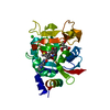
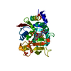
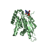
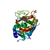
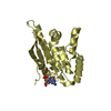
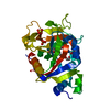
- PDB-6nri: Crystal Structure of human PARP-1 ART domain bound to inhibitor UTT83 -
+ Open data

- Basic information
| Entry | Database: PDB / ID: 6nri | ||||||
|---|---|---|---|---|---|---|---|
| Title | Crystal Structure of human PARP-1 ART domain bound to inhibitor UTT83 | ||||||
 Components Components | Poly [ADP-ribose] polymerase 1 | ||||||
 Keywords Keywords | transferase/transferase inhibitor /  PARP-1 / poly(ADP-ribose) polymerase / PARP inhibitor / PARP1 / ARTD1 / TRANSFERASE / transferase-transferase inhibitor complex PARP-1 / poly(ADP-ribose) polymerase / PARP inhibitor / PARP1 / ARTD1 / TRANSFERASE / transferase-transferase inhibitor complex | ||||||
| Function / homology |  Function and homology information Function and homology informationNAD+-histone H2BS6 serine ADP-ribosyltransferase activity / NAD+-histone H3S10 serine ADP-ribosyltransferase activity / NAD+-histone H2BE35 glutamate ADP-ribosyltransferase activity / regulation of base-excision repair / positive regulation of myofibroblast differentiation / NAD+-protein-tyrosine ADP-ribosyltransferase activity / NAD+-protein-histidine ADP-ribosyltransferase activity / negative regulation of ATP biosynthetic process / carbohydrate biosynthetic process / regulation of circadian sleep/wake cycle, non-REM sleep ...NAD+-histone H2BS6 serine ADP-ribosyltransferase activity / NAD+-histone H3S10 serine ADP-ribosyltransferase activity / NAD+-histone H2BE35 glutamate ADP-ribosyltransferase activity / regulation of base-excision repair / positive regulation of myofibroblast differentiation / NAD+-protein-tyrosine ADP-ribosyltransferase activity / NAD+-protein-histidine ADP-ribosyltransferase activity / negative regulation of ATP biosynthetic process / carbohydrate biosynthetic process / regulation of circadian sleep/wake cycle, non-REM sleep / positive regulation of single strand break repair / vRNA Synthesis / NAD+-protein-serine ADP-ribosyltransferase activity / negative regulation of adipose tissue development / NAD DNA ADP-ribosyltransferase activity / NAD+- protein-aspartate ADP-ribosyltransferase activity / NAD+-protein-glutamate ADP-ribosyltransferase activity / DNA ADP-ribosylation / mitochondrial DNA metabolic process / regulation of oxidative stress-induced neuron intrinsic apoptotic signaling pathway / signal transduction involved in regulation of gene expression / replication fork reversal / positive regulation of necroptotic process / HDR through MMEJ (alt-NHEJ) / ATP generation from poly-ADP-D-ribose / regulation of catalytic activity / transcription regulator activator activity / : / positive regulation of DNA-templated transcription, elongation / NAD+ ADP-ribosyltransferase / cellular response to zinc ion / negative regulation of telomere maintenance via telomere lengthening / positive regulation of intracellular estrogen receptor signaling pathway / protein auto-ADP-ribosylation / positive regulation of mitochondrial depolarization / response to aldosterone / mitochondrial DNA repair / negative regulation of cGAS/STING signaling pathway / positive regulation of double-strand break repair via homologous recombination / protein poly-ADP-ribosylation / positive regulation of cardiac muscle hypertrophy / nuclear replication fork / negative regulation of transcription elongation by RNA polymerase II / site of DNA damage / NAD+-protein ADP-ribosyltransferase activity / R-SMAD binding / positive regulation of SMAD protein signal transduction / macrophage differentiation / protein autoprocessing / decidualization / NAD+ ADP-ribosyltransferase activity / Transferases; Glycosyltransferases; Pentosyltransferases / nucleosome binding / POLB-Dependent Long Patch Base Excision Repair / SUMOylation of DNA damage response and repair proteins / protein localization to chromatin / mitochondrion organization / telomere maintenance / negative regulation of innate immune response / nucleotidyltransferase activity / cellular response to nerve growth factor stimulus / transforming growth factor beta receptor signaling pathway / nuclear estrogen receptor binding / protein-DNA complex / response to gamma radiation / Downregulation of SMAD2/3:SMAD4 transcriptional activity / DNA Damage Recognition in GG-NER / protein modification process / Dual Incision in GG-NER / Formation of Incision Complex in GG-NER / positive regulation of protein localization to nucleus / histone deacetylase binding / cellular response to insulin stimulus / cellular response to amyloid-beta / cellular response to UV / NAD binding / regulation of protein localization / double-strand break repair / site of double-strand break / cellular response to oxidative stress / nuclear envelope / positive regulation of canonical NF-kappaB signal transduction / transcription regulator complex / RNA polymerase II-specific DNA-binding transcription factor binding / transcription by RNA polymerase II / chromosome, telomeric region / damaged DNA binding / nuclear body / DNA repair / innate immune response / negative regulation of DNA-templated transcription / apoptotic process / DNA damage response / chromatin binding / ubiquitin protein ligase binding / chromatin / nucleolus / protein kinase binding / enzyme binding / negative regulation of transcription by RNA polymerase IISimilarity search - Function | ||||||
| Biological species |  Homo sapiens (human) Homo sapiens (human) | ||||||
| Method | X-RAY DIFFRACTION / SYNCHROTRON / MOLECULAR REPLACEMENT / molecular replacement / Resolution: 2.2 Å | ||||||
 Authors Authors | Langelier, M.F. / Pascal, J.M. | ||||||
| Funding support |  Canada, 1items Canada, 1items
| ||||||
 Citation Citation | Journal: J.Med.Chem. / Year: 2019 Title: Design and Synthesis of Poly(ADP-ribose) Polymerase Inhibitors: Impact of Adenosine Pocket-Binding Motif Appendage to the 3-Oxo-2,3-dihydrobenzofuran-7-carboxamide on Potency and Selectivity. Authors: Velagapudi, U.K. / Langelier, M.F. / Delgado-Martin, C. / Diolaiti, M.E. / Bakker, S. / Ashworth, A. / Patel, B.A. / Shao, X. / Pascal, J.M. / Talele, T.T. | ||||||
| History |
|
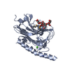
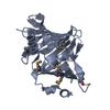
- Structure visualization
| Structure viewer | Molecule: MolmilJmol/JSmol |
|---|
- Downloads & links
-Download
| PDBx/mmCIF format | 6nri.cif.gz | 116.6 KB | Display | PDBx/mmCIF format |
|---|---|---|---|---|
| PDB format | pdb6nri.ent.gz | 87.3 KB | Display | PDB format |
| PDBx/mmJSON format | 6nri.json.gz | Tree view | PDBx/mmJSON format | |
| Others |  Other downloads Other downloads |
-Validation report
| Arichive directory | https://data.pdbj.org/pub/pdb/validation_reports/nr/6nriftp://data.pdbj.org/pub/pdb/validation_reports/nr/6nri | HTTPS FTP |
|---|
-Related structure data
| Related structure data |  6nrfC 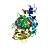 6nrgC  6nrhC  6nrjC  6bhvS C: citing same article ( S: Starting model for refinement |
|---|---|
| Similar structure data |

-Links
 PDBj
PDBj


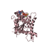
- Assembly
| Deposited unit | 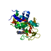
| ||||||||
|---|---|---|---|---|---|---|---|---|---|
| 1 |
| ||||||||
| Unit cell |
|
-Components
-Protein , 1 types, 1 molecules A
| #1: Protein | Mass: 30003.275 Da / Num. of mol.: 1 / Fragment: ADP-ribosyltransferase (ART) domain Source method: isolated from a genetically manipulated source Source: (gene. exp.) Homo sapiens (human) / Gene: PARP1, ADPRT, PPOL / Plasmid: pET28 / Production host:  Escherichia coli (E. coli) / Strain (production host): Rosetta2(DE3) Escherichia coli (E. coli) / Strain (production host): Rosetta2(DE3)References: UniProt: P09874, NAD+ ADP-ribosyltransferase, Transferases; Glycosyltransferases; Pentosyltransferases |
|---|
-Non-polymers , 5 types, 60 molecules 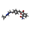


| #2: Chemical | ChemComp-KYM / ( Mass: 455.465 Da / Num. of mol.: 1 / Source method: obtained synthetically / Formula: C25H21N5O4 Mass: 455.465 Da / Num. of mol.: 1 / Source method: obtained synthetically / Formula: C25H21N5O4 |
|---|---|
| #3: Chemical | ChemComp-CIT / Citric acid Mass: 192.124 Da / Num. of mol.: 1 / Source method: obtained synthetically / Formula: C6H8O7 Mass: 192.124 Da / Num. of mol.: 1 / Source method: obtained synthetically / Formula: C6H8O7 |
| #4: Chemical | ChemComp-DMS / Dimethyl sulfoxide Mass: 78.133 Da / Num. of mol.: 1 / Source method: obtained synthetically / Formula: C2H6OS / Comment: DMSO, precipitant*YM Mass: 78.133 Da / Num. of mol.: 1 / Source method: obtained synthetically / Formula: C2H6OS / Comment: DMSO, precipitant*YM |
| #5: Chemical | ChemComp-CL / Chloride Mass: 35.453 Da / Num. of mol.: 1 / Source method: obtained synthetically / Formula: Cl Mass: 35.453 Da / Num. of mol.: 1 / Source method: obtained synthetically / Formula: Cl |
| #6: Water | ChemComp-HOH / WaterMass: 18.015 Da / Num. of mol.: 56 / Source method: isolated from a natural source / Formula: H2O |
-Experimental details
-Experiment
| Experiment | Method: X-RAY DIFFRACTION / Number of used crystals: 1 |
|---|
- Sample preparation
| Crystal | Density Matthews: 2.54 Å3/Da / Density % sol: 51.55 % / Mosaicity: 0.17 ° |
|---|---|
| Crystal grow | Temperature: 298 K / Method: evaporation / pH: 7.5 Details: ~20% PEG 3350, 0.2 M ammonium sulfate or sodium citrate, 100 mM Hepes pH 7.5 |
-Data collection
| Diffraction | Mean temperature: 100 K / Serial crystal experiment: N | ||||||||||||||||||||||||
|---|---|---|---|---|---|---|---|---|---|---|---|---|---|---|---|---|---|---|---|---|---|---|---|---|---|
| Diffraction source | Source: SYNCHROTRON / Site: CLSI / Beamline: 08ID-1 / Wavelength: 0.979 Å | ||||||||||||||||||||||||
| Detector | Type: DECTRIS PILATUS3 S 6M / Detector: PIXEL / Date: Feb 8, 2018 | ||||||||||||||||||||||||
| Radiation | Monochromator: ACCEL/BRUKER double crystal monochromator (DCM), Si(111) Protocol: SINGLE WAVELENGTH / Monochromatic (M) / Laue (L): M / Scattering type: x-ray | ||||||||||||||||||||||||
| Radiation wavelength | Wavelength: 0.979 Å / Relative weight: 1 | ||||||||||||||||||||||||
| Reflection | Resolution: 2.2→48.2 Å / Num. obs: 16019 / % possible obs: 99.9 % / Redundancy: 15.8 % / CC1/2: 0.999 / Rmerge(I) obs: 0.081 / Rpim(I) all: 0.021 / Rrim(I) all: 0.084 / Net I/σ(I): 20.3 | ||||||||||||||||||||||||
| Reflection shell | Diffraction-ID: 1
|
-Phasing
| Phasing | Method: molecular replacement |
|---|
- Processing
| Software |
| ||||||||||||||||||||||||||||||||||||||||||||||||||||||||||||
|---|---|---|---|---|---|---|---|---|---|---|---|---|---|---|---|---|---|---|---|---|---|---|---|---|---|---|---|---|---|---|---|---|---|---|---|---|---|---|---|---|---|---|---|---|---|---|---|---|---|---|---|---|---|---|---|---|---|---|---|---|---|
| Refinement | Method to determine structure: MOLECULAR REPLACEMENT Starting model: PDBID 6BHV Resolution: 2.2→48.2 Å / Cor.coef. Fo:Fc: 0.968 / Cor.coef. Fo:Fc free: 0.947 / SU B: 11.229 / SU ML: 0.139 / SU R Cruickshank DPI: 0.2204 / Cross valid method: THROUGHOUT / σ(F): 0 / ESU R: 0.22 / ESU R Free: 0.194 Details: HYDROGENS HAVE BEEN ADDED IN THE RIDING POSITIONS U VALUES : WITH TLS ADDED
| ||||||||||||||||||||||||||||||||||||||||||||||||||||||||||||
| Solvent computation | Ion probe radii: 0.7 Å / Shrinkage radii: 0.7 Å / VDW probe radii: 1 Å | ||||||||||||||||||||||||||||||||||||||||||||||||||||||||||||
| Displacement parameters | Biso max: 118.3 Å2 / Biso mean: 56.441 Å2 / Biso min: 37.57 Å2
| ||||||||||||||||||||||||||||||||||||||||||||||||||||||||||||
| Refinement step | Cycle: final / Resolution: 2.2→48.2 Å
| ||||||||||||||||||||||||||||||||||||||||||||||||||||||||||||
| Refine LS restraints |
| ||||||||||||||||||||||||||||||||||||||||||||||||||||||||||||
| LS refinement shell | Resolution: 2.202→2.259 Å / Rfactor Rfree error: 0 / Total num. of bins used: 20
| ||||||||||||||||||||||||||||||||||||||||||||||||||||||||||||
| Refinement TLS params. | Method: refined / Origin x: 25.1952 Å / Origin y: -4.5335 Å / Origin z: 20.062 Å
|
