 Movie
Movie Controller

[English] 日本語
 Yorodumi
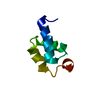
- PDB-6nl7: Crystal structure of B1 immunoglobulin-binding domain of Streptoc... -
+ Open data

- Basic information
| Entry | Database: PDB / ID: 6nl7 | ||||||
|---|---|---|---|---|---|---|---|
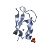
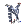
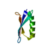
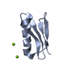
| Title | Crystal structure of B1 immunoglobulin-binding domain of Streptococcal Protein G (T16F, T18A, V21H, T25H, K28Y, V29I, K31R, Q32A, Y33L, N35K, D36A, N37Q) | ||||||
 Components Components | Immunoglobulin G-binding protein G | ||||||
 Keywords Keywords | METAL BINDING PROTEIN / Metal-mediated complex / B1 Domain of Streptococcal protein G /  Immunoglobulin binding protein Immunoglobulin binding protein | ||||||
| Function / homology |  Function and homology information Function and homology information | ||||||
| Biological species |  Streptococcus (bacteria) Streptococcus (bacteria) | ||||||
| Method | X-RAY DIFFRACTION / SYNCHROTRON / MOLECULAR REPLACEMENT / Resolution: 1.4 Å | ||||||
 Authors Authors | Maniaci, B. / Stec, B. / Huxford, T. | ||||||
 Citation Citation | Journal: Biochemistry / Year: 2019 Title: Design of High-Affinity Metal-Controlled Protein Dimers. Authors: Maniaci, B. / Lipper, C.H. / Anipindi, D.L. / Erlandsen, H. / Cole, J.L. / Stec, B. / Huxford, T. / Love, J.J. | ||||||
| History |
|
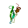
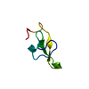
- Structure visualization
| Structure viewer | Molecule: MolmilJmol/JSmol |
|---|
- Downloads & links
-Download
| PDBx/mmCIF format | 6nl7.cif.gz | 133.3 KB | Display | PDBx/mmCIF format |
|---|---|---|---|---|
| PDB format | pdb6nl7.ent.gz | 103 KB | Display | PDB format |
| PDBx/mmJSON format | 6nl7.json.gz | Tree view | PDBx/mmJSON format | |
| Others |  Other downloads Other downloads |
-Validation report
| Arichive directory | https://data.pdbj.org/pub/pdb/validation_reports/nl/6nl7ftp://data.pdbj.org/pub/pdb/validation_reports/nl/6nl7 | HTTPS FTP |
|---|
-Related structure data
| Related structure data | 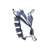 6nl6C 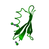 6nl8C 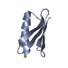 6nl9C 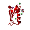 6nlaC  6nlbC 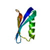 1pgaS S: Starting model for refinement C: citing same article ( |
|---|---|
| Similar structure data |


-Links
 PDBj
PDBj

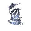
- Assembly
| Deposited unit | 
| ||||||||
|---|---|---|---|---|---|---|---|---|---|
| 1 | 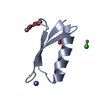
| ||||||||
| 2 | 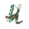
| ||||||||
| 3 | 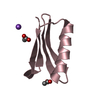
| ||||||||
| 4 | 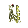
| ||||||||
| Unit cell |
|
-Components
-Antibody , 1 types, 4 molecules ABCD
| #1: Antibody | Mass: 6247.947 Da / Num. of mol.: 4 Mutation: T16F, T18A, V21H, T25H, K28Y, V29I, K31R, Q32A, Y33L, N35K, D36A, N37Q Source method: isolated from a genetically manipulated source Source: (gene. exp.) Streptococcus (bacteria) / Gene: spg / Plasmid: pet21a / Production host: Escherichia coli BL21(DE3) (bacteria) / Strain (production host): BL21(DE3) / References: UniProt: P19909 |
|---|
-Non-polymers , 7 types, 355 molecules 


| #2: Chemical | ChemComp-ZN /  Mass: 65.409 Da / Num. of mol.: 4 / Source method: obtained synthetically / Formula: Zn Mass: 65.409 Da / Num. of mol.: 4 / Source method: obtained synthetically / Formula: Zn#3: Chemical | ChemComp-ACT / Acetate Mass: 59.044 Da / Num. of mol.: 8 / Source method: obtained synthetically / Formula: C2H3O2 Mass: 59.044 Da / Num. of mol.: 8 / Source method: obtained synthetically / Formula: C2H3O2#4: Chemical | ChemComp-NA /  Mass: 22.990 Da / Num. of mol.: 4 / Source method: obtained synthetically / Formula: Na Mass: 22.990 Da / Num. of mol.: 4 / Source method: obtained synthetically / Formula: Na#5: Chemical | ChemComp-CL / | Chloride Mass: 35.453 Da / Num. of mol.: 1 / Source method: obtained synthetically / Formula: Cl Mass: 35.453 Da / Num. of mol.: 1 / Source method: obtained synthetically / Formula: Cl#6: Chemical | Phosphate Mass: 94.971 Da / Num. of mol.: 2 / Source method: isolated from a natural source / Formula: PO4 Mass: 94.971 Da / Num. of mol.: 2 / Source method: isolated from a natural source / Formula: PO4#7: Chemical | ChemComp-DPO / | Pyrophosphate Mass: 173.943 Da / Num. of mol.: 1 / Source method: obtained synthetically / Formula: O7P2 Mass: 173.943 Da / Num. of mol.: 1 / Source method: obtained synthetically / Formula: O7P2#8: Water | ChemComp-HOH / | WaterMass: 18.015 Da / Num. of mol.: 335 / Source method: isolated from a natural source / Formula: H2O |
|---|
-Experimental details
-Experiment
| Experiment | Method: X-RAY DIFFRACTION / Number of used crystals: 1 |
|---|
- Sample preparation
| Crystal | Density Matthews: 2.67 Å3/Da / Density % sol: 53.98 % |
|---|---|
| Crystal grow | Temperature: 298 K / Method: vapor diffusion, hanging drop Details: 80 mM acetic acid pH 3.6, 20 mM acetic acid pH 5.8, 30% 2,4-methylpentanediol, 200 mM NaCl and 20 mM zinc sulfate PH range: 3.6 - 5.8 |
-Data collection
| Diffraction | Mean temperature: 100 K / Serial crystal experiment: N |
|---|---|
| Diffraction source | Source: SYNCHROTRON / Site: ALS  / Beamline: 8.2.2 / Wavelength: 1.098 Å / Beamline: 8.2.2 / Wavelength: 1.098 Å |
| Detector | Type: ADSC QUANTUM 315r / Detector: CCD / Date: May 5, 2018 |
| Radiation | Protocol: SINGLE WAVELENGTH / Monochromatic (M) / Laue (L): M / Scattering type: x-ray |
| Radiation wavelength | Wavelength: 1.098 Å / Relative weight: 1 |
| Reflection | Resolution: 1.4→51.8 Å / Num. obs: 44547 / % possible obs: 90.03 % / Redundancy: 9.9 % / Rmerge(I) obs: 0.1 / Net I/σ(I): 7.8 |
| Reflection shell | Resolution: 1.398→1.434 Å / Num. unique obs: 932 / % possible all: 25.89 |
- Processing
| Software |
| ||||||||||||||||||||||||||||||||||||||||||||||||||||||||||||||||||||||||||||||||||||||||||||||||||||||||||||||||||||||||||||||||||||||||||||||||||||||||||||||||||||||||||||||||||||||
|---|---|---|---|---|---|---|---|---|---|---|---|---|---|---|---|---|---|---|---|---|---|---|---|---|---|---|---|---|---|---|---|---|---|---|---|---|---|---|---|---|---|---|---|---|---|---|---|---|---|---|---|---|---|---|---|---|---|---|---|---|---|---|---|---|---|---|---|---|---|---|---|---|---|---|---|---|---|---|---|---|---|---|---|---|---|---|---|---|---|---|---|---|---|---|---|---|---|---|---|---|---|---|---|---|---|---|---|---|---|---|---|---|---|---|---|---|---|---|---|---|---|---|---|---|---|---|---|---|---|---|---|---|---|---|---|---|---|---|---|---|---|---|---|---|---|---|---|---|---|---|---|---|---|---|---|---|---|---|---|---|---|---|---|---|---|---|---|---|---|---|---|---|---|---|---|---|---|---|---|---|---|---|---|
| Refinement | Method to determine structure: MOLECULAR REPLACEMENT Starting model: 1PGA Resolution: 1.4→40.06 Å / Cor.coef. Fo:Fc: 0.981 / Cor.coef. Fo:Fc free: 0.967 / SU B: 1.647 / SU ML: 0.029 / Cross valid method: THROUGHOUT / ESU R: 0.052 / ESU R Free: 0.053 / Stereochemistry target values: MAXIMUM LIKELIHOOD / Details: HYDROGENS HAVE BEEN ADDED IN THE RIDING POSITIONS
| ||||||||||||||||||||||||||||||||||||||||||||||||||||||||||||||||||||||||||||||||||||||||||||||||||||||||||||||||||||||||||||||||||||||||||||||||||||||||||||||||||||||||||||||||||||||
| Solvent computation | Ion probe radii: 0.8 Å / Shrinkage radii: 0.8 Å / VDW probe radii: 1.2 Å / Solvent model: MASK | ||||||||||||||||||||||||||||||||||||||||||||||||||||||||||||||||||||||||||||||||||||||||||||||||||||||||||||||||||||||||||||||||||||||||||||||||||||||||||||||||||||||||||||||||||||||
| Displacement parameters | Biso mean: 19.862 Å2
| ||||||||||||||||||||||||||||||||||||||||||||||||||||||||||||||||||||||||||||||||||||||||||||||||||||||||||||||||||||||||||||||||||||||||||||||||||||||||||||||||||||||||||||||||||||||
| Refinement step | Cycle: 1 / Resolution: 1.4→40.06 Å
| ||||||||||||||||||||||||||||||||||||||||||||||||||||||||||||||||||||||||||||||||||||||||||||||||||||||||||||||||||||||||||||||||||||||||||||||||||||||||||||||||||||||||||||||||||||||
| Refine LS restraints |
|