 Movie
Movie Controller

[English] 日本語
 Yorodumi
- PDB-6net: FAD-dependent monooxygenase TropB from T. stipitatus substrate complex -
+ Open data

- Basic information
| Entry | Database: PDB / ID: 6net | |||||||||
|---|---|---|---|---|---|---|---|---|---|---|
| Title | FAD-dependent monooxygenase TropB from T. stipitatus substrate complex | |||||||||
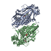
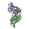
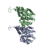
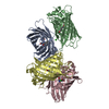
 Components Components | FAD-dependent monooxygenase tropB | |||||||||
 Keywords Keywords |  FLAVOPROTEIN / oxidative dearomatization FLAVOPROTEIN / oxidative dearomatization | |||||||||
| Function / homology |  Function and homology information Function and homology information | |||||||||
| Biological species | Talaromyces stipitatus | |||||||||
| Method | X-RAY DIFFRACTION / SYNCHROTRON / MOLECULAR REPLACEMENT / Resolution: 2.25 Å | |||||||||
 Authors Authors | Rodriguez Benitez, A. / Tweedy, S.E. / Baker Dockrey, S.A. / Lukowski, A.L. / Wymore, T. / Khare, D. / Brooks, C.L. / Palfey, B.A. / Smith, J.L. / Narayan, A.R.H. | |||||||||
| Funding support |  United States, 2items United States, 2items
| |||||||||
 Citation Citation | Journal: Acs Catalysis / Year: 2019 Title: Structural basis for selectivity in flavin-dependent monooxygenase-catalyzed oxidative dearomatization. Authors: Rodriguez Benitez, A. / Tweedy, S.E. / Baker Dockrey, S.A. / Lukowski, A.L. / Wymore, T. / Khare, D. / Brooks 3rd, C.L. / Palfey, B.A. / Smith, J.L. / Narayan, A.R.H. | |||||||||
| History |
|
- Structure visualization
| Structure viewer | Molecule: MolmilJmol/JSmol |
|---|
- Downloads & links
-Download
| PDBx/mmCIF format | 6net.cif.gz | 410.9 KB | Display | PDBx/mmCIF format |
|---|---|---|---|---|
| PDB format | pdb6net.ent.gz | 275.2 KB | Display | PDB format |
| PDBx/mmJSON format | 6net.json.gz | Tree view | PDBx/mmJSON format | |
| Others |  Other downloads Other downloads |
-Validation report
| Arichive directory | https://data.pdbj.org/pub/pdb/validation_reports/ne/6netftp://data.pdbj.org/pub/pdb/validation_reports/ne/6net | HTTPS FTP |
|---|
-Related structure data
| Related structure data |  6nesSC  6neuC  6nevC S: Starting model for refinement C: citing same article ( |
|---|---|
| Similar structure data |


-Links
 PDBj
PDBj- Assembly
| Deposited unit | 
| ||||||||||||
|---|---|---|---|---|---|---|---|---|---|---|---|---|---|
| 1 |
| ||||||||||||
| 2 | 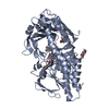
| ||||||||||||
| 3 | 
| ||||||||||||
| Unit cell |
|
-Components
-Protein , 1 types, 2 molecules AB
| #1: Protein | Mass: 49941.824 Da / Num. of mol.: 2 Source method: isolated from a genetically manipulated source Source: (gene. exp.)  Talaromyces stipitatus (strain ATCC 10500 / CBS 375.48 / QM 6759 / NRRL 1006) (fungus) Talaromyces stipitatus (strain ATCC 10500 / CBS 375.48 / QM 6759 / NRRL 1006) (fungus)Strain: ATCC 10500 / CBS 375.48 / QM 6759 / NRRL 1006 / Gene: tropB, tsL1, TSTA_117740 / Production host:  Escherichia coli (E. coli) / References: UniProt: B8M9J8 Escherichia coli (E. coli) / References: UniProt: B8M9J8 |
|---|
-Non-polymers , 5 types, 133 molecules 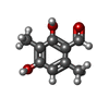


| #2: Chemical |  Mass: 166.174 Da / Num. of mol.: 3 / Source method: obtained synthetically / Formula: C9H10O3 Mass: 166.174 Da / Num. of mol.: 3 / Source method: obtained synthetically / Formula: C9H10O3#3: Chemical | Flavin adenine dinucleotide Mass: 785.550 Da / Num. of mol.: 2 / Source method: obtained synthetically / Formula: C27H33N9O15P2 / Feature type: SUBJECT OF INVESTIGATION / Comment: FAD*YM Mass: 785.550 Da / Num. of mol.: 2 / Source method: obtained synthetically / Formula: C27H33N9O15P2 / Feature type: SUBJECT OF INVESTIGATION / Comment: FAD*YM#4: Chemical | Chloride Mass: 35.453 Da / Num. of mol.: 2 / Source method: obtained synthetically / Formula: Cl / Feature type: SUBJECT OF INVESTIGATION Mass: 35.453 Da / Num. of mol.: 2 / Source method: obtained synthetically / Formula: Cl / Feature type: SUBJECT OF INVESTIGATION#5: Chemical | ChemComp-GOL / | Glycerol Mass: 92.094 Da / Num. of mol.: 1 / Source method: obtained synthetically / Formula: C3H8O3 Mass: 92.094 Da / Num. of mol.: 1 / Source method: obtained synthetically / Formula: C3H8O3#6: Water | ChemComp-HOH / | WaterMass: 18.015 Da / Num. of mol.: 125 / Source method: isolated from a natural source / Formula: H2O |
|---|
-Experimental details
-Experiment
| Experiment | Method: X-RAY DIFFRACTION / Number of used crystals: 1 |
|---|
- Sample preparation
| Crystal | Density Matthews: 2.78 Å3/Da / Density % sol: 55.81 % |
|---|---|
| Crystal grow | Temperature: 293.15 K / Method: vapor diffusion, sitting drop / pH: 7.8 / Details: 1.11 M ammonium tartrate dibasic, 6% hexanediol |
-Data collection
| Diffraction | Mean temperature: 100 K / Serial crystal experiment: N |
|---|---|
| Diffraction source | Source: SYNCHROTRON / Site: APS / Beamline: 23-ID-B / Wavelength: 1.033 Å |
| Detector | Type: DECTRIS EIGER X 16M / Detector: PIXEL / Date: Apr 9, 2018 |
| Radiation | Protocol: SINGLE WAVELENGTH / Monochromatic (M) / Laue (L): M / Scattering type: x-ray |
| Radiation wavelength | Wavelength: 1.033 Å / Relative weight: 1 |
| Reflection | Resolution: 2.245→45.96 Å / Num. obs: 47286 / % possible obs: 99.8 % / Redundancy: 13.3 % / Biso Wilson estimate: 51.95 Å2 / CC1/2: 0.996 / Rmerge(I) obs: 0.19 / Net I/σ(I): 1 |
| Reflection shell | Resolution: 2.245→2.325 Å / Rmerge(I) obs: 2.457 / CC1/2: 0.41 |
- Processing
| Software |
| ||||||||||||||||||||||||||||||||||||||||||||||||||||||||||||||||||||||||||||||||||||||||||||||||||||||||||||||||||||||||||||||||||||||||||||||||||||||||||||||||||||||||||||||||||||||||||||||||||||
|---|---|---|---|---|---|---|---|---|---|---|---|---|---|---|---|---|---|---|---|---|---|---|---|---|---|---|---|---|---|---|---|---|---|---|---|---|---|---|---|---|---|---|---|---|---|---|---|---|---|---|---|---|---|---|---|---|---|---|---|---|---|---|---|---|---|---|---|---|---|---|---|---|---|---|---|---|---|---|---|---|---|---|---|---|---|---|---|---|---|---|---|---|---|---|---|---|---|---|---|---|---|---|---|---|---|---|---|---|---|---|---|---|---|---|---|---|---|---|---|---|---|---|---|---|---|---|---|---|---|---|---|---|---|---|---|---|---|---|---|---|---|---|---|---|---|---|---|---|---|---|---|---|---|---|---|---|---|---|---|---|---|---|---|---|---|---|---|---|---|---|---|---|---|---|---|---|---|---|---|---|---|---|---|---|---|---|---|---|---|---|---|---|---|---|---|---|---|
| Refinement | Method to determine structure: MOLECULAR REPLACEMENT Starting model: 6NES Resolution: 2.25→45.96 Å / SU ML: 0.3754 / Cross valid method: FREE R-VALUE / σ(F): 1.35 / Phase error: 28.2893
| ||||||||||||||||||||||||||||||||||||||||||||||||||||||||||||||||||||||||||||||||||||||||||||||||||||||||||||||||||||||||||||||||||||||||||||||||||||||||||||||||||||||||||||||||||||||||||||||||||||
| Solvent computation | Shrinkage radii: 0.9 Å / VDW probe radii: 1.11 Å | ||||||||||||||||||||||||||||||||||||||||||||||||||||||||||||||||||||||||||||||||||||||||||||||||||||||||||||||||||||||||||||||||||||||||||||||||||||||||||||||||||||||||||||||||||||||||||||||||||||
| Displacement parameters | Biso mean: 62.05 Å2 | ||||||||||||||||||||||||||||||||||||||||||||||||||||||||||||||||||||||||||||||||||||||||||||||||||||||||||||||||||||||||||||||||||||||||||||||||||||||||||||||||||||||||||||||||||||||||||||||||||||
| Refinement step | Cycle: LAST / Resolution: 2.25→45.96 Å
| ||||||||||||||||||||||||||||||||||||||||||||||||||||||||||||||||||||||||||||||||||||||||||||||||||||||||||||||||||||||||||||||||||||||||||||||||||||||||||||||||||||||||||||||||||||||||||||||||||||
| Refine LS restraints |
| ||||||||||||||||||||||||||||||||||||||||||||||||||||||||||||||||||||||||||||||||||||||||||||||||||||||||||||||||||||||||||||||||||||||||||||||||||||||||||||||||||||||||||||||||||||||||||||||||||||
| LS refinement shell |
|
