[English] 日本語
Yorodumi
- PDB-6mvq: HCV NS5B 1b N316 bound to Compound 31 -

+
Open data


ID or keywords:

Loading...

-
Basic information

Entry
Database: PDB / ID: 6mvq
TitleHCV NS5B 1b N316 bound to Compound 31
ComponentsHCV Polymerase
Keywords
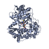
VIRAL PROTEIN / Hepatitus C / NS5B / REPLICATION
Function / homology
Function and homology information


host cell mitochondrial membrane / host cell lipid droplet / symbiont-mediated suppression of host TRAF-mediated signal transduction / transformation of host cell by virus / symbiont-mediated perturbation of host cell cycle G1/S transition checkpoint / symbiont-mediated suppression of host JAK-STAT cascade via inhibition of STAT1 activity / symbiont-mediated suppression of host cytoplasmic pattern recognition receptor signaling pathway via inhibition of MAVS activity / ribonucleoside triphosphate phosphatase activity / lipid droplet / : ...host cell mitochondrial membrane / host cell lipid droplet / symbiont-mediated suppression of host TRAF-mediated signal transduction / transformation of host cell by virus / symbiont-mediated perturbation of host cell cycle G1/S transition checkpoint / symbiont-mediated suppression of host JAK-STAT cascade via inhibition of STAT1 activity / symbiont-mediated suppression of host cytoplasmic pattern recognition receptor signaling pathway via inhibition of MAVS activity / ribonucleoside triphosphate phosphatase activity / lipid droplet / : / protein complex oligomerization / monoatomic ion channel activity / clathrin-dependent endocytosis of virus by host cell / viral nucleocapsid / RNA helicase activity / host cell endoplasmic reticulum membrane / host cell perinuclear region of cytoplasm / induction by virus of host autophagy / ribonucleoprotein complex / viral RNA genome replication / cysteine-type endopeptidase activity / RNA-dependent RNA polymerase activity / serine-type endopeptidase activity / fusion of virus membrane with host endosome membrane / viral envelope / symbiont-mediated suppression of host type I interferon-mediated signaling pathway / structural molecule activity / virion attachment to host cell / host cell plasma membrane / virion membrane / proteolysis / RNA binding / zinc ion binding / ATP binding / plasma membrane / cytoplasm
Similarity search - Function
Hepatitis C virus core protein, chain A superfamily / Hepatitus C virus, Non-structural 5a protein, C-terminal / Hepatitis C virus NS5A, 1B domain superfamily / Hepatitis C virus non-structural protein NS2, C-terminal domain / Hepatitis C virus non-structural protein NS2, N-terminal domain / Hepatitis C virus non-structural protein NS2 / HCV NS5a protein C-terminal region / Hepatitis C virus, Non-structural protein NS4b / Hepatitis C virus, Core protein, N-terminal / Hepatitis C virus non-structural protein NS4b ...Hepatitis C virus core protein, chain A superfamily / Hepatitus C virus, Non-structural 5a protein, C-terminal / Hepatitis C virus NS5A, 1B domain superfamily / Hepatitis C virus non-structural protein NS2, C-terminal domain / Hepatitis C virus non-structural protein NS2, N-terminal domain / Hepatitis C virus non-structural protein NS2 / HCV NS5a protein C-terminal region / Hepatitis C virus, Non-structural protein NS4b / Hepatitis C virus, Core protein, N-terminal / Hepatitis C virus non-structural protein NS4b / Hepatitis C virus capsid protein / Hepatitis C virus, Non-structural protein NS2 / Hepatitis C virus, Non-structural 5a protein / Hepatitis C virus, Non-structural 5a protein, domain 1a / Hepatitis C virus non-structural 5a, 1B domain / NS5A domain 1a superfamily / Hepatitis C virus non-structural 5a protein membrane anchor / Hepatitis C virus non-structural 5a zinc finger domain / Hepatitis C virus non-structural 5a domain 1b / Hepacivirus nonstructural protein 2 (NS2) protease domain profile. / Hepatitis C virus, Non-structural protein NS4a / Hepatitis C virus non-structural protein NS4a / Hepatitis C virus, Core protein, C-terminal / Hepatitis C virus core protein / Hepatitis C virus, Non-structural protein E2/NS1 / Hepatitis C virus non-structural protein E2/NS1 / Hepatitis C virus, Envelope glycoprotein E1 / Hepatitis C virus envelope glycoprotein E1 / RNA dependent RNA polymerase, hepatitis C virus / Viral RNA dependent RNA polymerase / Hepatitis C virus, NS3 protease, Peptidase S29 / Hepatitis C virus NS3 protease / Hepacivirus/Pegivirus NS3 protease domain profile. / Reverse transcriptase/Diguanylate cyclase domain / DEAD box, Flavivirus / Flavivirus DEAD domain / Superfamilies 1 and 2 helicase C-terminal domain profile. / Superfamilies 1 and 2 helicase ATP-binding type-1 domain profile. / DEAD-like helicases superfamily / Helicase, C-terminal / Helicase superfamily 1/2, ATP-binding domain / Reverse transcriptase/Diguanylate cyclase domain / RNA-directed RNA polymerase, catalytic domain / RdRp of positive ssRNA viruses catalytic domain profile. / Alpha-Beta Plaits / Peptidase S1, PA clan, chymotrypsin-like fold / Peptidase S1, PA clan / DNA/RNA polymerase superfamily / P-loop containing nucleoside triphosphate hydrolase / 2-Layer Sandwich / Alpha Beta
Similarity search - Domain/homology
Chem-K4M / Genome polyprotein
Similarity search - Component
Biological speciesHepatitis C virus subtype 1b
MethodX-RAY DIFFRACTION / SYNCHROTRON / MOLECULAR REPLACEMENT / molecular replacement / Resolution: 2.14 Å
Model detailsHCV NS5B 1b N316 bound to Compound 31
AuthorsWilliams, S.P. / Kahler, K. / Price, D.J. / Peat, A.J.
Citation
Journal: J.Med.Chem. / Year: 2019
Title: Design of N-Benzoxaborole Benzofuran GSK8175-Optimization of Human Pharmacokinetics Inspired by Metabolites of a Failed Clinical HCV Inhibitor.
Authors: Chong, P.Y. / Shotwell, J.B. / Miller, J. / Price, D.J. / Maynard, A. / Voitenleitner, C. / Mathis, A. / Williams, S. / Pouliot, J.J. / Creech, K. / Wang, F. / Fang, J. / Zhang, H. / Tai, V. ...Authors: Chong, P.Y. / Shotwell, J.B. / Miller, J. / Price, D.J. / Maynard, A. / Voitenleitner, C. / Mathis, A. / Williams, S. / Pouliot, J.J. / Creech, K. / Wang, F. / Fang, J. / Zhang, H. / Tai, V.W. / Turner, E. / Kahler, K.M. / Crosby, R. / Peat, A.J.
#1: Journal: J. Med. Chem. / Year: 2014
Title: Discovery of a potent boronic acid derived inhibitor of the HCV RNA-dependent RNA polymerase.
Authors: Maynard, A. / Crosby, R.M. / Ellis, B. / Hamatake, R. / Hong, Z. / Johns, B.A. / Kahler, K.M. / Koble, C. / Leivers, A. / Leivers, M.R. / Mathis, A. / Peat, A.J. / Pouliot, J.J. / Roberts, C. ...Authors: Maynard, A. / Crosby, R.M. / Ellis, B. / Hamatake, R. / Hong, Z. / Johns, B.A. / Kahler, K.M. / Koble, C. / Leivers, A. / Leivers, M.R. / Mathis, A. / Peat, A.J. / Pouliot, J.J. / Roberts, C.D. / Samano, V. / Schmidt, R.M. / Smith, G.K. / Spaltenstein, A. / Stewart, E.L. / Thommes, P. / Turner, E.M. / Voitenleitner, C. / Walker, J.T. / Waitt, G. / Weatherhead, J. / Weaver, K. / Williams, S. / Wright, L. / Xiong, Z.Z. / Haigh, D. / Shotwell, J.B.
History
DepositionOct 26, 2018Deposition site: RCSB / Processing site: RCSB
Revision 1.0Sep 4, 2019Provider: repository / Type: Initial release
Revision 1.1Mar 13, 2024Group: Data collection / Database references / Category: chem_comp_atom / chem_comp_bond / database_2
Item: _database_2.pdbx_DOI / _database_2.pdbx_database_accession

-
Structure visualization

Structure viewerMolecule:
MolmilJmol/JSmol

Downloads & links

-
Assembly

Deposited unit
A: HCV Polymerase
B: HCV Polymerase
hetero molecules


Theoretical massNumber of molelcules
Total (without water)125,9864
Polymers124,9572
Non-polymers1,0292
Water8,035446
1
A: HCV Polymerase
hetero molecules


Theoretical massNumber of molelcules
Total (without water)62,9932
Polymers62,4791
Non-polymers5141
Water181
TypeNameSymmetry operationNumber
identity operation1_555x,y,z1
MethodPISA
2
B: HCV Polymerase
hetero molecules


Theoretical massNumber of molelcules
Total (without water)62,9932
Polymers62,4791
Non-polymers5141
Water181
TypeNameSymmetry operationNumber
identity operation1_555x,y,z1
MethodPISA
Unit cell
Length a, b, c (Å)85.811, 106.565, 126.509
Angle α, β, γ (deg.)90.000, 90.000, 90.000
Int Tables number19
Space group name H-MP212121

-
Components

#1: Protein HCV Polymerase


Mass: 62478.605 Da / Num. of mol.: 2 / Mutation: F101Y, K114R
Source method: isolated from a genetically manipulated source
Source: (gene. exp.) Hepatitis C virus subtype 1b / Plasmid: pET28 / Production host: Escherichia coli (E. coli) / Strain (production host): BL21(DE3) / References: UniProt: Q99AU2
#2: Chemical ChemComp-K4M / (4-{1-[5-cyclopropyl-2-(4-fluorophenyl)-3-(methylcarbamoyl)-1-benzofuran-6-yl]-1H-1,2,4-triazol-5-yl}-2-fluorophenyl)boronic acid


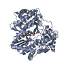
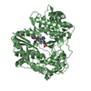
Mass: 514.288 Da / Num. of mol.: 2 / Source method: obtained synthetically / Formula: C27H21BF2N4O4
#3: Water ChemComp-HOH / water / Water


Mass: 18.015 Da / Num. of mol.: 446 / Source method: isolated from a natural source / Formula: H2O

-
Experimental details

-
Experiment

ExperimentMethod: X-RAY DIFFRACTION / Number of used crystals: 1

-
Sample preparation

CrystalDensity Matthews: 2.31 Å3/Da / Density % sol: 46.86 % / Mosaicity: 0.991 °
Crystal growTemperature: 298 K / Method: vapor diffusion, hanging drop / pH: 5 / Details: 0.1M Na Citrate pH5.0, 10% glycerol, 15% PEG3350

-
Data collection

DiffractionMean temperature: 100 K / Serial crystal experiment: N
Diffraction sourceSource: SYNCHROTRON / Site: APS / Beamline: 21-ID-F / Wavelength: 0.987 Å
DetectorType: RAYONIX MX-300 / Detector: CCD / Date: Aug 23, 2011
RadiationProtocol: SINGLE WAVELENGTH / Monochromatic (M) / Laue (L): M / Scattering type: x-ray
Radiation wavelengthWavelength: 0.987 Å / Relative weight: 1
ReflectionResolution: 2.07→50 Å / Num. obs: 72869 / % possible obs: 99.6 % / Redundancy: 6 % / Rmerge(I) obs: 0.088 / Χ2: 1 / Net I/σ(I): 5.7
Reflection shell
Resolution (Å)Redundancy (%)Rmerge(I) obsNum. unique obsΧ2Diffraction-ID% possible all
2.07-2.1460.59971600.847199.3
2.14-2.2360.46172010.861199.4
2.23-2.3360.3871610.903199.5
2.33-2.4560.28572250.931199.5
2.45-2.616.10.21272260.929199.6
2.61-2.816.10.15572600.944199.8
2.81-3.096.10.11272721.025199.9
3.09-3.546.10.07573401.118199.9
3.54-4.466.10.04973901.13199.9
4.46-505.80.04876341.299199.5

-
Phasing

PhasingMethod: molecular replacement

-
Processing

Software
NameVersionClassificationNB
HKL-2000data reduction
HKL-2000data scaling
MOLREPphasing
REFMAC5.6.0081refinement
PDB_EXTRACT3.24data extraction
RefinementMethod to determine structure: MOLECULAR REPLACEMENT / Resolution: 2.14→29.68 Å / Cor.coef. Fo:Fc: 0.957 / Cor.coef. Fo:Fc free: 0.945 / SU B: 9.9 / SU ML: 0.118 / SU R Cruickshank DPI: 0.0596 / Cross valid method: THROUGHOUT / σ(F): 0 / ESU R: 0.06 / ESU R Free: 0.042
Details: U VALUES : WITH TLS ADDED HYDROGENS HAVE BEEN USED IF PRESENT IN THE INPUT
RfactorNum. reflection% reflectionSelection details
Rfree0.2376 4659 7.2 %RANDOM
Rwork0.2103 ---
obs0.2123 59756 99.6 %-
Solvent computationIon probe radii: 0.8 Å / Shrinkage radii: 0.8 Å / VDW probe radii: 1.2 Å
Displacement parametersBiso max: 71.09 Å2 / Biso mean: 34.16 Å2 / Biso min: 22.32 Å2
Baniso -1Baniso -2Baniso -3
1--41.46 Å20 Å20 Å2
2--53.8 Å20 Å2
3----12.35 Å2
Refinement stepCycle: final / Resolution: 2.14→29.68 Å
ProteinNucleic acidLigandSolventTotal
Num. atoms8439 0 76 446 8961
Biso mean--38.26 41.42 -
Num. residues----1105
Refine LS restraints
Refine-IDTypeDev idealDev ideal targetNumber
X-RAY DIFFRACTIONr_bond_refined_d0.0080.0228735
X-RAY DIFFRACTIONr_bond_other_d0.0020.025802
X-RAY DIFFRACTIONr_angle_refined_deg1.1361.97111907
X-RAY DIFFRACTIONr_angle_other_deg1.117314146
X-RAY DIFFRACTIONr_dihedral_angle_1_deg5.08751111
X-RAY DIFFRACTIONr_dihedral_angle_2_deg31.89823.097339
X-RAY DIFFRACTIONr_dihedral_angle_3_deg12.303151400
X-RAY DIFFRACTIONr_dihedral_angle_4_deg12.9741558
X-RAY DIFFRACTIONr_chiral_restr0.0510.21358
X-RAY DIFFRACTIONr_gen_planes_refined0.0040.0219718
X-RAY DIFFRACTIONr_gen_planes_other0.0020.021768
Refine LS restraints NCSNCS model details: RESTRAINTS / Rms dev Biso : 0 Å2 / Rms dev position: 0 Å / Weight Biso : 0 / Weight position: 0
LS refinement shellResolution: 2.139→2.194 Å / Rfactor Rfree error: 0 / Total num. of bins used: 20
RfactorNum. reflection% reflection
Rfree0.295 331 -
Rwork0.255 4240 -
all-4571 -
obs--97.65 %
Refinement TLS params.

Method: refined / Refine-ID: X-RAY DIFFRACTION

IDL112)L122)L132)L222)L232)L332)S11 (Å °)S12 (Å °)S13 (Å °)S21 (Å °)S22 (Å °)S23 (Å °)S31 (Å °)S32 (Å °)S33 (Å °)T112)T122)T132)T222)T232)T332)Origin x (Å)Origin y (Å)Origin z (Å)
10.2403-0.08390.28210.0441-0.11160.64270.038-0.01620.0005-0.0102-0.0017-0.0050.0477-0.0202-0.03630.3202-0.0198-0.00770.00490.00350.309311.26845.90647.254
20.25910.04690.13270.03740.03260.36870.02170.0347-0.00090.0106-0.00050.00670.02550.0336-0.02120.31830.0129-0.0060.0074-0.00230.318-11.36245.915-7.925
Refinement TLS group
IDRefine-IDRefine TLS-IDAuth asym-IDAuth seq-ID
1X-RAY DIFFRACTION1A1 - 563
2X-RAY DIFFRACTION1A601
3X-RAY DIFFRACTION2B1 - 563
4X-RAY DIFFRACTION2B601

+
About Yorodumi

-
News

-
Feb 9, 2022. New format data for meta-information of EMDB entries

New format data for meta-information of EMDB entries

  • Version 3 of the EMDB header file is now the official format.
  • The previous official version 1.9 will be removed from the archive.

Related info.:EMDB header

External links:wwPDB to switch to version 3 of the EMDB data model

-
Aug 12, 2020. Covid-19 info

Covid-19 info

URL: https://pdbj.org/emnavi/covid19.php

New page: Covid-19 featured information page in EM Navigator.

Related info.:Covid-19 info / Mar 5, 2020. Novel coronavirus structure data

+
Mar 5, 2020. Novel coronavirus structure data

Novel coronavirus structure data

Related info.:Yorodumi Speices / Aug 12, 2020. Covid-19 info

External links:COVID-19 featured content - PDBj / Molecule of the Month (242):Coronavirus Proteases

+
Jan 31, 2019. EMDB accession codes are about to change! (news from PDBe EMDB page)

EMDB accession codes are about to change! (news from PDBe EMDB page)

  • The allocation of 4 digits for EMDB accession codes will soon come to an end. Whilst these codes will remain in use, new EMDB accession codes will include an additional digit and will expand incrementally as the available range of codes is exhausted. The current 4-digit format prefixed with “EMD-” (i.e. EMD-XXXX) will advance to a 5-digit format (i.e. EMD-XXXXX), and so on. It is currently estimated that the 4-digit codes will be depleted around Spring 2019, at which point the 5-digit format will come into force.
  • The EM Navigator/Yorodumi systems omit the EMD- prefix.

Related info.:Q: What is EMD? / ID/Accession-code notation in Yorodumi/EM Navigator

External links:EMDB Accession Codes are Changing Soon! / Contact to PDBj

+
Jul 12, 2017. Major update of PDB

Major update of PDB

  • wwPDB released updated PDB data conforming to the new PDBx/mmCIF dictionary.
  • This is a major update changing the version number from 4 to 5, and with Remediation, in which all the entries are updated.
  • In this update, many items about electron microscopy experimental information are reorganized (e.g. em_software).
  • Now, EM Navigator and Yorodumi are based on the updated data.

External links:wwPDB Remediation / Enriched Model Files Conforming to OneDep Data Standards Now Available in the PDB FTP Archive

-
Yorodumi

Thousand views of thousand structures

  • Yorodumi is a browser for structure data from EMDB, PDB, SASBDB, etc.
  • This page is also the successor to EM Navigator detail page, and also detail information page/front-end page for Omokage search.
  • The word "yorodu" (or yorozu) is an old Japanese word meaning "ten thousand". "mi" (miru) is to see.

Related info.:EMDB / PDB / SASBDB / Comparison of 3 databanks / Yorodumi Search / Aug 31, 2016. New EM Navigator & Yorodumi / Yorodumi Papers / Jmol/JSmol / Function and homology information / Changes in new EM Navigator and Yorodumi

Read more