 Movie
Movie Controller

[English] 日本語
 Yorodumi
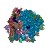
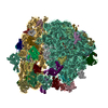
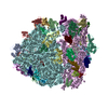
- PDB-6lkq: The Structural Basis for Inhibition of Ribosomal Translocation by... -
+ Open data

- Basic information
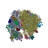
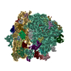
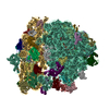
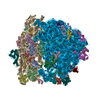
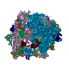
| Entry | Database: PDB / ID: 6lkq | ||||||
|---|---|---|---|---|---|---|---|
| Title | The Structural Basis for Inhibition of Ribosomal Translocation by Viomycin | ||||||
 Components Components |
| ||||||
 Keywords Keywords |  TRANSLATION / 70S ribosome viomycin translocation TRANSLATION / 70S ribosome viomycin translocation | ||||||
| Function / homology |  Function and homology information Function and homology informationregulation of translational termination / translation release factor activity, codon specific / ribosomal large subunit assembly / large ribosomal subunit rRNA binding / large ribosomal subunit / small ribosomal subunit / 5S rRNA binding / transferase activity / tRNA binding / rRNA binding ...regulation of translational termination / translation release factor activity, codon specific / ribosomal large subunit assembly / large ribosomal subunit rRNA binding / large ribosomal subunit / small ribosomal subunit / 5S rRNA binding / transferase activity / tRNA binding / rRNA binding / ribosome / structural constituent of ribosome / translation / ribonucleoprotein complex / mRNA binding / GTPase activity / GTP binding / cytosol / cytoplasmSimilarity search - Function | ||||||
| Biological species |  Escherichia coli (E. coli) Escherichia coli (E. coli)synthetic construct (others) | ||||||
| Method | X-RAY DIFFRACTION / SYNCHROTRON / MOLECULAR REPLACEMENT / Resolution: 3.1 Å | ||||||
 Authors Authors | Zhang, L. / Wang, Y.H. / Lancaster, L. / Zhou, J. / Noller, H.F. | ||||||
| Funding support |  China, 1items China, 1items
| ||||||
 Citation Citation | Journal: Proc Natl Acad Sci U S A / Year: 2020 Title: The structural basis for inhibition of ribosomal translocation by viomycin. Authors: Ling Zhang / Ying-Hui Wang / Xing Zhang / Laura Lancaster / Jie Zhou / Harry F Noller /  Abstract: Viomycin, an antibiotic that has been used to fight tuberculosis infections, is believed to block the translocation step of protein synthesis by inhibiting ribosomal subunit dissociation and trapping ...Viomycin, an antibiotic that has been used to fight tuberculosis infections, is believed to block the translocation step of protein synthesis by inhibiting ribosomal subunit dissociation and trapping the ribosome in an intermediate state of intersubunit rotation. The mechanism by which viomycin stabilizes this state remains unexplained. To address this, we have determined cryo-EM and X-ray crystal structures of 70S ribosome complexes trapped in a rotated state by viomycin. The 3.8-Å resolution cryo-EM structure reveals a ribosome trapped in the hybrid state with 8.6° intersubunit rotation and 5.3° rotation of the 30S subunit head domain, bearing a single P/E state transfer RNA (tRNA). We identify five different binding sites for viomycin, four of which have not been previously described. To resolve the details of their binding interactions, we solved the 3.1-Å crystal structure of a viomycin-bound ribosome complex, revealing that all five viomycins bind to ribosomal RNA. One of these (Vio1) corresponds to the single viomycin that was previously identified in a complex with a nonrotated classical-state ribosome. Three of the newly observed binding sites (Vio3, Vio4, and Vio5) are clustered at intersubunit bridges, consistent with the ability of viomycin to inhibit subunit dissociation. We propose that one or more of these same three viomycins induce intersubunit rotation by selectively binding the rotated state of the ribosome at dynamic elements of 16S and 23S rRNA, thus, blocking conformational changes associated with molecular movements that are required for translocation. | ||||||
| History |
|
- Structure visualization
| Structure viewer | Molecule: MolmilJmol/JSmol |
|---|
- Downloads & links
-Download
| PDBx/mmCIF format | 6lkq.cif.gz | 3.6 MB | Display | PDBx/mmCIF format |
|---|---|---|---|---|
| PDB format | pdb6lkq.ent.gz | Display | PDB format | |
| PDBx/mmJSON format | 6lkq.json.gz | Tree view | PDBx/mmJSON format | |
| Others |  Other downloads Other downloads |
-Validation report
| Arichive directory | https://data.pdbj.org/pub/pdb/validation_reports/lk/6lkqftp://data.pdbj.org/pub/pdb/validation_reports/lk/6lkq | HTTPS FTP |
|---|
-Related structure data
| Related structure data | 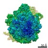 0939C  3f1e 3f1f S: Starting model for refinement C: citing same article ( |
|---|---|
| Similar structure data |


-Links
 PDBj
PDBj


- Assembly
| Deposited unit | 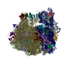
| ||||||||
|---|---|---|---|---|---|---|---|---|---|
| 1 |
| ||||||||
| Unit cell |
|
-Components
-30S ribosomal protein ... , 20 types, 20 molecules ABCDEFGHIJKLMNOPQRST
| #1: Protein | Mass: 24253.943 Da / Num. of mol.: 1 / Source method: isolated from a natural source / Source: (natural) Escherichia coli (E. coli) / Strain: MRE600 / References: UniProt: C3TPN2 |
|---|---|
| #2: Protein | Mass: 23078.785 Da / Num. of mol.: 1 / Source method: isolated from a natural source / Source: (natural) Escherichia coli (E. coli) / Strain: MRE600 / References: UniProt: A0A376HTV6 |
| #3: Protein | Mass: 23383.002 Da / Num. of mol.: 1 / Source method: isolated from a natural source / Source: (natural) Escherichia coli (E. coli) / Strain: MRE600 / References: UniProt: A0A4P8BV83 |
| #4: Protein | Mass: 15804.282 Da / Num. of mol.: 1 / Source method: isolated from a natural source / Source: (natural) Escherichia coli (E. coli) / Strain: MRE600 / References: UniProt: F4SQ30 |
| #5: Protein | Mass: 11669.371 Da / Num. of mol.: 1 / Source method: isolated from a natural source / Source: (natural) Escherichia coli (E. coli) / Strain: MRE600 / References: UniProt: A0A3L2GT12, UniProt: A0A7J9U446*PLUS |
| #6: Protein | Mass: 16861.523 Da / Num. of mol.: 1 / Source method: isolated from a natural source / Source: (natural) Escherichia coli (E. coli) / Strain: MRE600 / References: UniProt: D6I218 |
| #7: Protein | Mass: 14015.361 Da / Num. of mol.: 1 / Source method: isolated from a natural source / Source: (natural) Escherichia coli (E. coli) / Strain: MRE600 / References: UniProt: U9ZUM7 |
| #8: Protein | Mass: 14554.882 Da / Num. of mol.: 1 / Source method: isolated from a natural source / Source: (natural) Escherichia coli (E. coli) / Strain: MRE600 / References: UniProt: D8A919 |
| #9: Protein | Mass: 11196.988 Da / Num. of mol.: 1 / Source method: isolated from a natural source / Source: (natural) Escherichia coli (E. coli) / Strain: MRE600 / References: UniProt: A0A5B9AU26 |
| #10: Protein | Mass: 12487.200 Da / Num. of mol.: 1 / Source method: isolated from a natural source / Source: (natural) Escherichia coli (E. coli) / Strain: MRE600 / References: UniProt: A0A4P8B3R1 |
| #11: Protein | Mass: 13636.961 Da / Num. of mol.: 1 / Source method: isolated from a natural source / Source: (natural) Escherichia coli (E. coli) / Strain: MRE600 / References: UniProt: L3C3P4 |
| #12: Protein | Mass: 12625.753 Da / Num. of mol.: 1 / Source method: isolated from a natural source / Source: (natural) Escherichia coli (E. coli) / Strain: MRE600 / References: UniProt: A0A069XLE9 |
| #13: Protein | Mass: 11489.390 Da / Num. of mol.: 1 / Source method: isolated from a natural source / Source: (natural) Escherichia coli (E. coli) / Strain: MRE600 / References: UniProt: A0A090BZT4 |
| #14: Protein | Mass: 10159.621 Da / Num. of mol.: 1 / Source method: isolated from a natural source / Source: (natural) Escherichia coli (E. coli) / Strain: MRE600 / References: UniProt: A0A029IK47 |
| #15: Protein | Mass: 9207.572 Da / Num. of mol.: 1 / Source method: isolated from a natural source / Source: (natural) Escherichia coli (E. coli) / Strain: MRE600 / References: UniProt: A0A029IMB2 |
| #16: Protein | Mass: 9263.946 Da / Num. of mol.: 1 / Source method: isolated from a natural source / Source: (natural) Escherichia coli (E. coli) / Strain: MRE600 / References: UniProt: T6LV72 |
| #17: Protein | Mass: 6466.477 Da / Num. of mol.: 1 / Source method: isolated from a natural source / Source: (natural) Escherichia coli (E. coli) / Strain: MRE600 / References: UniProt: E9TDX6 |
| #18: Protein | Mass: 9057.626 Da / Num. of mol.: 1 / Source method: isolated from a natural source / Source: (natural) Escherichia coli (E. coli) / Strain: MRE600 / References: UniProt: S1EA57 |
| #19: Protein | Mass: 9506.190 Da / Num. of mol.: 1 / Source method: isolated from a natural source / Source: (natural) Escherichia coli (E. coli) / Strain: MRE600 / References: UniProt: D7ZAS2 |
| #20: Protein | Mass: 6067.081 Da / Num. of mol.: 1 / Source method: isolated from a natural source / Source: (natural) Escherichia coli (E. coli) / Strain: MRE600 / References: UniProt: A0A5C9AJ78 |
+50S ribosomal protein ... , 30 types, 33 molecules UVWXYZ012345689abcdefghijklmno...
-RNA chain , 4 types, 4 molecules stuw
| #51: RNA chain | Mass: 496563.125 Da / Num. of mol.: 1 / Source method: isolated from a natural source / Source: (natural) Escherichia coli (E. coli) / Strain: MRE600 / References: GenBank: 1726022446 |
|---|---|
| #52: RNA chain | Mass: 941306.188 Da / Num. of mol.: 1 / Source method: isolated from a natural source / Source: (natural) Escherichia coli (E. coli) / Strain: MRE 600 / References: GenBank: 1036415628 |
| #53: RNA chain | Mass: 38177.762 Da / Num. of mol.: 1 / Source method: isolated from a natural source / Source: (natural) Escherichia coli (E. coli) / Strain: MRE600 / References: GenBank: 1727529157 |
| #55: RNA chain | Mass: 1900.198 Da / Num. of mol.: 1 / Source method: isolated from a natural source / Source: (natural) Escherichia coli (E. coli) |
-Protein / Protein/peptide , 2 types, 6 molecules vyz7AABA
| #54: Protein | Mass: 59152.379 Da / Num. of mol.: 1 / Source method: isolated from a natural source / Source: (natural) Escherichia coli (E. coli) / References: UniProt: C3SE77 |
|---|---|
| #56: Protein/peptide | / 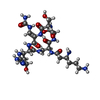  Type: Oligopeptide / Class: Antibiotic / Mass: 703.707 Da / Num. of mol.: 5 / Source method: obtained synthetically / Source: (synth.) synthetic construct (others) / References: Viomycin Type: Oligopeptide / Class: Antibiotic / Mass: 703.707 Da / Num. of mol.: 5 / Source method: obtained synthetically / Source: (synth.) synthetic construct (others) / References: Viomycin |
-Details
| Has ligand of interest | Y |
|---|
-Experimental details
-Experiment
| Experiment | Method: X-RAY DIFFRACTION / Number of used crystals: 1 |
|---|
- Sample preparation
| Crystal | Density Matthews: 3.04 Å3/Da / Density % sol: 59.59 % |
|---|---|
| Crystal grow | Temperature: 295 K / Method: vapor diffusion, sitting drop Details: Tris Ac PH.7.0, 25-35 mM KCL, 6.1% PEG 20000, 1% glycerol, 50mM sucrose |
-Data collection
| Diffraction | Mean temperature: 100 K / Serial crystal experiment: N |
|---|---|
| Diffraction source | Source: SYNCHROTRON / Site: APS / Beamline: 23-ID-D / Wavelength: 1.03318 Å |
| Detector | Type: MAR scanner 300 mm plate / Detector: IMAGE PLATE / Date: Apr 2, 2011 |
| Radiation | Protocol: SINGLE WAVELENGTH / Monochromatic (M) / Laue (L): M / Scattering type: x-ray |
| Radiation wavelength | Wavelength: 1.03318 Å / Relative weight: 1 |
| Reflection | Resolution: 3.1→100 Å / Num. obs: 433307 / % possible obs: 100 % / Redundancy: 7 % / Rmerge(I) obs: 0.2 / Rsym value: 0.1 / Net I/σ(I): 1.7 |
| Reflection shell | Resolution: 3.1→50 Å / Num. unique obs: 433307 / CC1/2: 0.6 |
- Processing
| Software |
| ||||||||||||||||||
|---|---|---|---|---|---|---|---|---|---|---|---|---|---|---|---|---|---|---|---|
| Refinement | Method to determine structure: MOLECULAR REPLACEMENT Starting model: 3F1E. 3F1F Resolution: 3.1→100 Å / Cross valid method: THROUGHOUT
| ||||||||||||||||||
| Displacement parameters | Biso max: 442.55 Å2 / Biso mean: 100.1107 Å2 / Biso min: 18.64 Å2 | ||||||||||||||||||
| Refinement step | Cycle: LAST / Resolution: 3.1→100 Å
| ||||||||||||||||||
| LS refinement shell | Resolution: 3.1→50 Å / Rfactor Rfree: 0.21 / Rfactor Rwork: 0.24 |
