Movie
Movie Controller


+ Open data

- Basic information
| Entry | Database: PDB / ID: 6k3i | |||||||||||||||
|---|---|---|---|---|---|---|---|---|---|---|---|---|---|---|---|---|
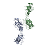
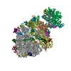
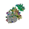
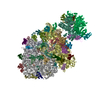
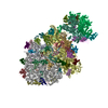
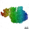
| Title | Salmonella hook in curved state - 66 subunit models | |||||||||||||||
 Components Components | Flagellar hook protein FlgE | |||||||||||||||
 Keywords Keywords |  MOTOR PROTEIN / bacterial / hook / flexible joint / flagella MOTOR PROTEIN / bacterial / hook / flexible joint / flagella | |||||||||||||||
| Function / homology |  Function and homology information Function and homology informationbacterial-type flagellum basal body / bacterial-type flagellum-dependent swarming motility Similarity search - Function | |||||||||||||||
| Biological species |  Salmonella typhimurium (bacteria) Salmonella typhimurium (bacteria) | |||||||||||||||
| Method | ELECTRON MICROSCOPY / single particle reconstruction / cryo EM / Resolution: 2.86 Å | |||||||||||||||
 Authors Authors | Shibata, S. / Matsunami, H. / Wolf, M. / Aizawa, S. | |||||||||||||||
| Funding support |  Japan, 4items Japan, 4items
| |||||||||||||||
 Citation Citation | Journal: Nat Struct Mol Biol / Year: 2019 Title: Torque transmission mechanism of the curved bacterial flagellar hook revealed by cryo-EM. Authors: Satoshi Shibata / Hideyuki Matsunami / Shin-Ichi Aizawa / Matthias Wolf / Abstract: Bacterial locomotion by rotating flagella is achieved through the hook, which transmits torque from the motor to the filament. The hook is a tubular structure composed of a single type of protein, ...Bacterial locomotion by rotating flagella is achieved through the hook, which transmits torque from the motor to the filament. The hook is a tubular structure composed of a single type of protein, yet it adopts a curved shape. To perform its function, it must be simultaneously flexible and torsionally rigid. The molecular mechanism by which chemically identical subunits form such a dynamic structure is unknown. Here, we show the complete structure of the hook from Salmonella enterica in its supercoiled 'curved' state, at 2.9 Å resolution. Subunits in the curved hook are grouped into 11 distinctive conformations, each shared along 11 protofilaments. The domains of the elongated hook subunit behave as rigid bodies connected by two hinge regions. The reconstituted model demonstrates how identical subunits can dynamically change conformation by physical interactions while bending. These multiple subunit states contradict the two-state model, which is a key feature of flagellar polymorphism. | |||||||||||||||
| History |
|
- Structure visualization
| Movie |
Movie viewer |
|---|---|
| Structure viewer | Molecule: MolmilJmol/JSmol |
- Downloads & links
-Download
| PDBx/mmCIF format | 6k3i.cif.gz | 4 MB | Display | PDBx/mmCIF format |
|---|---|---|---|---|
| PDB format | pdb6k3i.ent.gz | Display | PDB format | |
| PDBx/mmJSON format | 6k3i.json.gz | Tree view | PDBx/mmJSON format | |
| Others |  Other downloads Other downloads |
-Validation report
| Arichive directory | https://data.pdbj.org/pub/pdb/validation_reports/k3/6k3iftp://data.pdbj.org/pub/pdb/validation_reports/k3/6k3i | HTTPS FTP |
|---|
-Related structure data
| Related structure data | 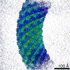 9909MC M: map data used to model this data C: citing same article ( |
|---|---|
| Similar structure data |

-Links
 PDBj
PDBj
- Assembly
| Deposited unit | 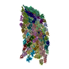
|
|---|---|
| 1 |
|
-Components
| #1: Protein | Mass: 42101.957 Da / Num. of mol.: 66 / Source method: isolated from a natural source Source: (natural) Salmonella typhimurium (strain LT2 / SGSC1412 / ATCC 700720) (bacteria)Strain: LT2 / SGSC1412 / ATCC 700720 / References: UniProt: P0A1J1 |
|---|
-Experimental details
-Experiment
| Experiment | Method: ELECTRON MICROSCOPY |
|---|---|
| EM experiment | Aggregation state: FILAMENT / 3D reconstruction method: single particle reconstruction |
- Sample preparation
| Component | Name: Bacterial flagellar polyhook / Type: COMPLEX / Entity ID: all / Source: NATURAL |
|---|---|
| Molecular weight | Value: 2.8 MDa / Experimental value: NO |
| Source (natural) | Organism: Salmonella enterica subsp. enterica serovar Typhimurium str. LT2 (bacteria) |
| Buffer solution | pH: 3.5 |
| Buffer component | Conc.: 50 mM / Name: Glycine / Formula: C2H5NO2 |
| Specimen | Embedding applied: NO / Shadowing applied: NO / Staining applied: NO / Vitrification applied: YES |
| Specimen support | Grid material: COPPER / Grid mesh size: 400 divisions/in. / Grid type: Quantifoil R1.2/1.3 |
| Vitrification | Instrument: FEI VITROBOT MARK IV / Cryogen name: ETHANE / Humidity: 100 % / Chamber temperature: 289 K / Details: 3 second blot, 4.0uL |
- Electron microscopy imaging
| Experimental equipment |  Model: Titan Krios / Image courtesy: FEI Company |
|---|---|
| Microscopy | Model: FEI TITAN KRIOS / Details: nanoprobe, parallel beam illumination |
| Electron gun | Electron source: FIELD EMISSION GUN / Accelerating voltage: 300 kV / Illumination mode: FLOOD BEAM |
| Electron lens | Mode: BRIGHT FIELDBright-field microscopy / Nominal magnification: 105000 X / Calibrated magnification: 107914 X / Nominal defocus max: 5130 nm / Nominal defocus min: 430 nm / Calibrated defocus min: 430 nm / Calibrated defocus max: 5130 nm / Cs: 2.7 mm / C2 aperture diameter: 50 µm / Alignment procedure: COMA FREE |
| Specimen holder | Cryogen: NITROGEN / Specimen holder model: FEI TITAN KRIOS AUTOGRID HOLDER / Temperature (max): 100 K / Temperature (min): 77 K |
| Image recording | Average exposure time: 2 sec. / Electron dose: 89 e/Å2 / Detector mode: INTEGRATING / Film or detector model: FEI FALCON III (4k x 4k) / Num. of grids imaged: 1 / Num. of real images: 2655 Details: frame alignment and dose weighting using motioncor2 |
| Image scans | Sampling size: 15 µm / Width: 4000 / Height: 4000 |
- Processing
| EM software |
| |||||||||||||||||||||||||||||||||||||||||||||
|---|---|---|---|---|---|---|---|---|---|---|---|---|---|---|---|---|---|---|---|---|---|---|---|---|---|---|---|---|---|---|---|---|---|---|---|---|---|---|---|---|---|---|---|---|---|---|
| Image processing | Details: frame alignment and integration with motioncor2 incl. dose weighting | |||||||||||||||||||||||||||||||||||||||||||||
| CTF correction | Details: deconvolution in cisTEM / Type: PHASE FLIPPING AND AMPLITUDE CORRECTION | |||||||||||||||||||||||||||||||||||||||||||||
| Particle selection | Num. of particles selected: 998061 | |||||||||||||||||||||||||||||||||||||||||||||
| Symmetry | Point symmetry: C1 (asymmetric) | |||||||||||||||||||||||||||||||||||||||||||||
| 3D reconstruction | Resolution: 2.86 Å / Resolution method: FSC 0.143 CUT-OFF / Num. of particles: 234536 / Algorithm: FOURIER SPACE / Num. of class averages: 1 / Symmetry type: POINT | |||||||||||||||||||||||||||||||||||||||||||||
| Atomic model building | Protocol: FLEXIBLE FIT / Space: REAL / Target criteria: CC | |||||||||||||||||||||||||||||||||||||||||||||
| Atomic model building | PDB-ID: 1WLG Pdb chain-ID: A / Accession code: 1WLG / Pdb chain residue range: 70-359 / Source name: PDB / Type: experimental model |