 Movie
Movie Controller


+ Open data

- Basic information
| Entry | Database: PDB / ID: 6jlq | |||||||||
|---|---|---|---|---|---|---|---|---|---|---|
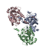
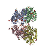
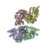
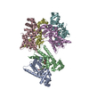
| Title | Crystal structure of human USP46-WDR48-WDR20 complex | |||||||||
 Components Components |
| |||||||||
 Keywords Keywords |  HYDROLASE / Deubiquitinase / DUB / USP46 HYDROLASE / Deubiquitinase / DUB / USP46 | |||||||||
| Function / homology |  Function and homology information Function and homology informationregulation of protein monoubiquitination / Signaling by cytosolic PDGFRA and PDGFRB fusion proteins / adult feeding behavior / righting reflex / behavioral response to ethanol / deubiquitinase activator activity / skeletal system morphogenesis / skin development / regulation of synaptic transmission, GABAergic / positive regulation of double-strand break repair via homologous recombination ...regulation of protein monoubiquitination / Signaling by cytosolic PDGFRA and PDGFRB fusion proteins / adult feeding behavior / righting reflex / behavioral response to ethanol / deubiquitinase activator activity / skeletal system morphogenesis / skin development / regulation of synaptic transmission, GABAergic / positive regulation of double-strand break repair via homologous recombination / seminiferous tubule development / protein deubiquitination / single fertilization / homeostasis of number of cells / behavioral fear response / embryonic organ development / ubiquitin binding / positive regulation of epithelial cell proliferation / Recognition of DNA damage by PCNA-containing replication complex / Fanconi Anemia Pathway / double-strand break repair via homologous recombination / positive regulation of receptor signaling pathway via JAK-STAT / multicellular organism growth / late endosome / single-stranded DNA binding / double-stranded DNA binding / spermatogenesis / ubiquitinyl hydrolase 1 / cysteine-type deubiquitinase activity / lysosome / Ub-specific processing proteases / intracellular membrane-bounded organelle / DNA damage response / proteolysis / DNA binding / nucleoplasm / metal ion binding / nucleus / cytosol / cytoplasmSimilarity search - Function | |||||||||
| Biological species |  Homo sapiens (human) Homo sapiens (human) Bos mutus (wild yak) Bos mutus (wild yak) | |||||||||
| Method | X-RAY DIFFRACTION / SYNCHROTRON / MOLECULAR REPLACEMENT / Resolution: 3.101 Å | |||||||||
 Authors Authors | Zhu, H. / Zhang, T. / Ding, J. | |||||||||
| Funding support |  China, 2items China, 2items
| |||||||||
 Citation Citation | Journal: Cell Discov / Year: 2019 Title: Structural insights into the activation of USP46 by WDR48 and WDR20. Authors: Zhu, H. / Zhang, T. / Wang, F. / Yang, J. / Ding, J. | |||||||||
| History |
|
- Structure visualization
| Structure viewer | Molecule: MolmilJmol/JSmol |
|---|
- Downloads & links
-Download
| PDBx/mmCIF format | 6jlq.cif.gz | 255.7 KB | Display | PDBx/mmCIF format |
|---|---|---|---|---|
| PDB format | pdb6jlq.ent.gz | 196.7 KB | Display | PDB format |
| PDBx/mmJSON format | 6jlq.json.gz | Tree view | PDBx/mmJSON format | |
| Others |  Other downloads Other downloads |
-Validation report
| Arichive directory | https://data.pdbj.org/pub/pdb/validation_reports/jl/6jlqftp://data.pdbj.org/pub/pdb/validation_reports/jl/6jlq | HTTPS FTP |
|---|
-Related structure data
| Related structure data | 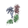 5k1cS S: Starting model for refinement |
|---|---|
| Similar structure data |


-Links
 PDBj
PDBj


- Assembly
| Deposited unit | 
| ||||||||
|---|---|---|---|---|---|---|---|---|---|
| 1 |
| ||||||||
| Unit cell |
|
-Components
-Protein , 1 types, 1 molecules A
| #1: Protein | Mass: 41262.852 Da / Num. of mol.: 1 Source method: isolated from a genetically manipulated source Source: (gene. exp.) Homo sapiens (human) / Gene: USP46 / Production host:  Escherichia coli (E. coli) / References: UniProt: P62068, ubiquitinyl hydrolase 1 Escherichia coli (E. coli) / References: UniProt: P62068, ubiquitinyl hydrolase 1 |
|---|
-WD repeat-containing protein ... , 2 types, 2 molecules BC
| #2: Protein | Mass: 69730.641 Da / Num. of mol.: 1 Source method: isolated from a genetically manipulated source Source: (gene. exp.) Homo sapiens (human) / Gene: WDR48 / Production host:   Spodoptera frugiperda (fall armyworm) / References: UniProt: Q8TAF3 Spodoptera frugiperda (fall armyworm) / References: UniProt: Q8TAF3 |
|---|---|
| #3: Protein | Mass: 48560.879 Da / Num. of mol.: 1 Fragment: UNP residues 1-318,UNP residues 106-137,UNP residues 535-595 Source method: isolated from a genetically manipulated source Source: (gene. exp.) Homo sapiens (human), (gene. exp.) Bos mutus (wild yak)Gene: WDR20, M91_05678 / Production host: Escherichia coli (E. coli)References: UniProt: Q8TBZ3, UniProt: B3KQX8, UniProt: L8I535 |
-Non-polymers , 3 types, 10 molecules 


| #4: Chemical | ChemComp-ZN /  Mass: 65.409 Da / Num. of mol.: 1 / Source method: obtained synthetically / Formula: Zn Mass: 65.409 Da / Num. of mol.: 1 / Source method: obtained synthetically / Formula: Zn | ||
|---|---|---|---|
| #5: Chemical | ChemComp-GOL / Glycerol Mass: 92.094 Da / Num. of mol.: 8 / Source method: obtained synthetically / Formula: C3H8O3 Mass: 92.094 Da / Num. of mol.: 8 / Source method: obtained synthetically / Formula: C3H8O3#6: Chemical | ChemComp-PO4 / | Phosphate Mass: 94.971 Da / Num. of mol.: 1 / Source method: obtained synthetically / Formula: PO4 Mass: 94.971 Da / Num. of mol.: 1 / Source method: obtained synthetically / Formula: PO4 |
-Experimental details
-Experiment
| Experiment | Method: X-RAY DIFFRACTION / Number of used crystals: 1 |
|---|
- Sample preparation
| Crystal | Density Matthews: 4.78 Å3/Da / Density % sol: 74.27 % |
|---|---|
| Crystal grow | Temperature: 289 K / Method: vapor diffusion, hanging drop Details: 1.0M Sodium phosphate monobasic monohydrate, Potassium phosphate dibasic, pH 7.2 |
-Data collection
| Diffraction | Mean temperature: 100 K / Serial crystal experiment: N | |||||||||||||||||||||||||||||||||||||||||||||||||||||||||||||||||||||||||||||||||||||||||||||||||||
|---|---|---|---|---|---|---|---|---|---|---|---|---|---|---|---|---|---|---|---|---|---|---|---|---|---|---|---|---|---|---|---|---|---|---|---|---|---|---|---|---|---|---|---|---|---|---|---|---|---|---|---|---|---|---|---|---|---|---|---|---|---|---|---|---|---|---|---|---|---|---|---|---|---|---|---|---|---|---|---|---|---|---|---|---|---|---|---|---|---|---|---|---|---|---|---|---|---|---|---|---|
| Diffraction source | Source: SYNCHROTRON / Site: SSRF / Beamline: BL17U1 / Wavelength: 0.9791 Å | |||||||||||||||||||||||||||||||||||||||||||||||||||||||||||||||||||||||||||||||||||||||||||||||||||
| Detector | Type: DECTRIS PILATUS 6M / Detector: PIXEL / Date: Oct 14, 2016 | |||||||||||||||||||||||||||||||||||||||||||||||||||||||||||||||||||||||||||||||||||||||||||||||||||
| Radiation | Protocol: SINGLE WAVELENGTH / Monochromatic (M) / Laue (L): M / Scattering type: x-ray | |||||||||||||||||||||||||||||||||||||||||||||||||||||||||||||||||||||||||||||||||||||||||||||||||||
| Radiation wavelength | Wavelength: 0.9791 Å / Relative weight: 1 | |||||||||||||||||||||||||||||||||||||||||||||||||||||||||||||||||||||||||||||||||||||||||||||||||||
| Reflection | Resolution: 3.1→50 Å / Num. obs: 56304 / % possible obs: 99 % / Redundancy: 11 % / Biso Wilson estimate: 88.06 Å2 / Rmerge(I) obs: 0.141 / Rpim(I) all: 0.041 / Rrim(I) all: 0.148 / Χ2: 2.519 / Net I/σ(I): 9.9 / Num. measured all: 616763 | |||||||||||||||||||||||||||||||||||||||||||||||||||||||||||||||||||||||||||||||||||||||||||||||||||
| Reflection shell | Diffraction-ID: 1
|
- Processing
| Software |
| |||||||||||||||||||||||||||||||||||||||||||||||||||||||||||||||||||||||||||||||||||||||||||||||||||||||||||||||||||||||||||||||||||||||||||||||||||
|---|---|---|---|---|---|---|---|---|---|---|---|---|---|---|---|---|---|---|---|---|---|---|---|---|---|---|---|---|---|---|---|---|---|---|---|---|---|---|---|---|---|---|---|---|---|---|---|---|---|---|---|---|---|---|---|---|---|---|---|---|---|---|---|---|---|---|---|---|---|---|---|---|---|---|---|---|---|---|---|---|---|---|---|---|---|---|---|---|---|---|---|---|---|---|---|---|---|---|---|---|---|---|---|---|---|---|---|---|---|---|---|---|---|---|---|---|---|---|---|---|---|---|---|---|---|---|---|---|---|---|---|---|---|---|---|---|---|---|---|---|---|---|---|---|---|---|---|---|
| Refinement | Method to determine structure: MOLECULAR REPLACEMENT Starting model: 5K1C Resolution: 3.101→43.986 Å / SU ML: 0.47 / Cross valid method: THROUGHOUT / σ(F): 1.35 / Phase error: 28.63
| |||||||||||||||||||||||||||||||||||||||||||||||||||||||||||||||||||||||||||||||||||||||||||||||||||||||||||||||||||||||||||||||||||||||||||||||||||
| Solvent computation | Shrinkage radii: 0.9 Å / VDW probe radii: 1.11 Å | |||||||||||||||||||||||||||||||||||||||||||||||||||||||||||||||||||||||||||||||||||||||||||||||||||||||||||||||||||||||||||||||||||||||||||||||||||
| Displacement parameters | Biso max: 183.11 Å2 / Biso mean: 95.0071 Å2 / Biso min: 30 Å2 | |||||||||||||||||||||||||||||||||||||||||||||||||||||||||||||||||||||||||||||||||||||||||||||||||||||||||||||||||||||||||||||||||||||||||||||||||||
| Refinement step | Cycle: final / Resolution: 3.101→43.986 Å
| |||||||||||||||||||||||||||||||||||||||||||||||||||||||||||||||||||||||||||||||||||||||||||||||||||||||||||||||||||||||||||||||||||||||||||||||||||
| Refine LS restraints |
| |||||||||||||||||||||||||||||||||||||||||||||||||||||||||||||||||||||||||||||||||||||||||||||||||||||||||||||||||||||||||||||||||||||||||||||||||||
| LS refinement shell | Refine-ID: X-RAY DIFFRACTION / Rfactor Rfree error: 0 / Total num. of bins used: 20
|
