[English] 日本語
Yorodumi
- PDB-6jgj: Crystal structure of the F99S/M153T/V163A/E222Q variant of GFP at... -

+
Open data


ID or keywords:

Loading...

-
Basic information

Entry
Database: PDB / ID: 6jgj
TitleCrystal structure of the F99S/M153T/V163A/E222Q variant of GFP at 0.78 A
ComponentsGreen fluorescent protein
KeywordsFLUORESCENT PROTEIN / Green Fluorescent Protein / hydrogen
Function / homology
Function and homology information


bioluminescence / generation of precursor metabolites and energy
Similarity search - Function
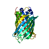
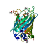
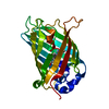
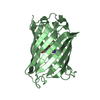
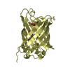
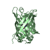
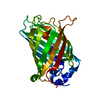
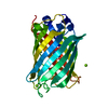
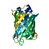
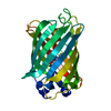
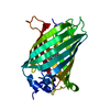
Green Fluorescent Protein / Green fluorescent protein / Green fluorescent protein, GFP / Green fluorescent protein-related / Green fluorescent protein / Green fluorescent protein / Beta Barrel / Mainly Beta
Similarity search - Domain/homology
Green fluorescent protein
Similarity search - Component
Biological speciesAequorea victoria (jellyfish)
MethodX-RAY DIFFRACTION / SYNCHROTRON / MOLECULAR REPLACEMENT / Resolution: 0.78 Å
AuthorsTakaba, K. / Tai, Y. / Hanazono, Y. / Miki, K. / Takeda, K.
Funding support Japan, 1items
OrganizationGrant numberCountry
Japan Society for the Promotion of Science17H03643 Japan
CitationJournal: Iucrj / Year: 2019
Title: Subatomic resolution X-ray structures of green fluorescent protein.
Authors: Takaba, K. / Tai, Y. / Eki, H. / Dao, H.A. / Hanazono, Y. / Hasegawa, K. / Miki, K. / Takeda, K.
History
DepositionFeb 14, 2019Deposition site: PDBJ / Processing site: PDBJ
Revision 1.0Apr 17, 2019Provider: repository / Type: Initial release
Revision 1.1May 1, 2019Group: Data collection / Database references / Category: pdbx_related_exp_data_set
Revision 1.2May 29, 2019Group: Data collection / Database references / Category: citation / citation_author
Item: _citation.page_first / _citation.page_last ..._citation.page_first / _citation.page_last / _citation.pdbx_database_id_PubMed / _citation.title / _citation_author.identifier_ORCID
Revision 2.0Nov 15, 2023Group: Atomic model / Data collection ...Atomic model / Data collection / Database references / Derived calculations
Category: atom_site / atom_site_anisotrop ...atom_site / atom_site_anisotrop / chem_comp_atom / chem_comp_bond / database_2 / pdbx_struct_conn_angle / struct_conn
Item: _atom_site.auth_atom_id / _atom_site.label_atom_id ..._atom_site.auth_atom_id / _atom_site.label_atom_id / _atom_site_anisotrop.pdbx_auth_atom_id / _atom_site_anisotrop.pdbx_label_atom_id / _database_2.pdbx_DOI / _database_2.pdbx_database_accession / _pdbx_struct_conn_angle.ptnr1_auth_seq_id / _pdbx_struct_conn_angle.ptnr1_label_alt_id / _pdbx_struct_conn_angle.ptnr1_symmetry / _pdbx_struct_conn_angle.ptnr3_auth_seq_id / _pdbx_struct_conn_angle.ptnr3_label_alt_id / _pdbx_struct_conn_angle.ptnr3_symmetry / _pdbx_struct_conn_angle.value / _struct_conn.pdbx_dist_value / _struct_conn.pdbx_leaving_atom_flag / _struct_conn.pdbx_ptnr1_label_alt_id / _struct_conn.pdbx_ptnr2_label_alt_id / _struct_conn.ptnr1_auth_seq_id / _struct_conn.ptnr1_label_asym_id / _struct_conn.ptnr1_label_atom_id / _struct_conn.ptnr2_auth_seq_id / _struct_conn.ptnr2_label_atom_id / _struct_conn.ptnr2_symmetry
Revision 2.1Nov 22, 2023Group: Refinement description / Category: pdbx_initial_refinement_model

-
Structure visualization

Structure viewerMolecule:
MolmilJmol/JSmol

Downloads & links

-
Assembly

Deposited unit
A: Green fluorescent protein
hetero molecules


Theoretical massNumber of molelcules
Total (without water)25,8923
Polymers25,8431
Non-polymers492
Water11,458636
1


  • Idetical with deposited unit
  • defined by author&software
TypeNameSymmetry operationNumber
identity operation1_555x,y,z1
Buried area40 Å2
ΔGint-2 kcal/mol
Surface area10380 Å2
MethodPISA
Unit cell
Length a, b, c (Å)50.870, 62.340, 68.850
Angle α, β, γ (deg.)90.000, 90.000, 90.000
Int Tables number19
Space group name H-MP212121

-
Components

#1: Protein Green fluorescent protein /


Mass: 25842.973 Da / Num. of mol.: 1 / Mutation: E222Q, F99S, M153T, V163A
Source method: isolated from a genetically manipulated source
Source: (gene. exp.) Aequorea victoria (jellyfish) / Gene: GFP / Plasmid: pET21a / Production host: Escherichia coli BL21(DE3) (bacteria) / Strain (production host): BL21(DE3) / References: UniProt: P42212
#2: Chemical ChemComp-MG / MAGNESIUM ION


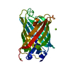
Mass: 24.305 Da / Num. of mol.: 2 / Source method: obtained synthetically / Formula: Mg
#3: Water ChemComp-HOH / water / Water


Mass: 18.015 Da / Num. of mol.: 636 / Source method: isolated from a natural source / Formula: H2O
Compound detailsThe authors state that there are some difference in the structures between CRO in database and CRO ...The authors state that there are some difference in the structures between CRO in database and CRO in this model. They are in the terminal carboxylic and amino groups and the protonation of phenolic oxygen.
Sequence details(1) The mutated chromophore, CRO, was used an initial model for refinement. Its hydroxyethyl group ...(1) The mutated chromophore, CRO, was used an initial model for refinement. Its hydroxyethyl group inserted to the model by S65T mutation was replaced with original hydoxymethyl in the refinement step, and finally its name was changed to GYS. (2) Q80R was caused by a PCR error in the early study (Chalfie, M. et al., Science, 1994).

-
Experimental details

-
Experiment

ExperimentMethod: X-RAY DIFFRACTION / Number of used crystals: 1

-
Sample preparation

CrystalDensity Matthews: 2.11 Å3/Da / Density % sol: 41.73 %
Crystal growTemperature: 308 K / Method: vapor diffusion / pH: 8.5 / Details: PEG 4000, MgCl2, Tris-HCl buffer

-
Data collection

DiffractionMean temperature: 56 K / Serial crystal experiment: N
Diffraction sourceSource: SYNCHROTRON / Site: SPring-8 / Beamline: BL41XU / Wavelength: 0.35 Å
DetectorType: DECTRIS PILATUS3 6M / Detector: PIXEL / Date: Jun 16, 2016
RadiationProtocol: SINGLE WAVELENGTH / Monochromatic (M) / Laue (L): M / Scattering type: x-ray
Radiation wavelengthWavelength: 0.35 Å / Relative weight: 1
ReflectionResolution: 0.77→31.14 Å / Num. obs: 254665 / % possible obs: 98.9 % / Observed criterion σ(I): -3 / Redundancy: 11.873 % / Biso Wilson estimate: 8.04 Å2 / CC1/2: 0.999 / Rmerge(I) obs: 0.073 / Rrim(I) all: 0.076 / Χ2: 0.949 / Net I/σ(I): 15.68
Reflection shell

Diffraction-ID: 1

Resolution (Å)Redundancy (%)Rmerge(I) obsMean I/σ(I) obsNum. unique obsCC1/2Rrim(I) all% possible all
0.78-0.88.632.0981.12227210.5072.22998
0.8-0.8111.031.711.56183730.6531.79399.8
0.81-0.8411.2821.3731.98179190.7341.43899.9
0.84-0.8611.4221.1532.35173750.791.20799.9
0.86-0.8911.5520.8883.09168940.8610.928100
0.89-0.9211.7020.6734.07163430.9090.704100
0.92-0.9611.860.4985.49157630.9490.52100
0.96-0.9912.0420.3677.3151910.9710.383100
0.99-1.0412.2230.2759.66145690.9850.287100
1.04-1.0912.4710.18513.71139330.9930.19399.9
1.09-1.1512.6720.12918.99133250.9960.135100
1.15-1.2212.9850.10622.64125880.9980.11100
1.22-1.312.9580.09425.01118220.9980.098100
1.3-1.4112.9640.08228.35110550.9980.08699.9
1.41-1.5413.4110.06434.67100220.9990.06798.4
1.54-1.7213.2530.04942.8489310.9990.05196.5
1.72-1.9911.2980.0447.7377600.9990.04294.5
1.99-2.4310.890.03656.4663820.9990.03791.4
2.43-3.4414.4150.03570.6754730.9990.037100
3.44-31.1413.0710.03971.7831270.9980.0499.7

-
Processing

Software
NameVersionClassification
SHELXLrefinement
XSCALEdata scaling
PDB_EXTRACT3.24data extraction
XDSdata reduction
PHASERphasing
RefinementMethod to determine structure: MOLECULAR REPLACEMENT
Starting model: 2WUR
Resolution: 0.78→31.14 Å / Cross valid method: FREE R-VALUE
Details: This model was finally refined with Multipolar Atomic Model using MoPro program but the determined multipolar parameters are not included.
RfactorNum. reflection% reflectionSelection details
Rfree0.125 12719 4.99 %Random selection
Rwork0.108 ---
obs-254639 99.2 %-
Displacement parametersBiso max: 85.31 Å2 / Biso mean: 11.0321 Å2 / Biso min: 4.57 Å2
Refinement stepCycle: LAST / Resolution: 0.78→31.14 Å
ProteinNucleic acidLigandSolventTotal
Num. atoms1803 0 1 636 2440
LS refinement shellResolution: 0.78→0.8 Å /
Num. reflection% reflection
obs22721 98 %

+
About Yorodumi

-
News

-
Feb 9, 2022. New format data for meta-information of EMDB entries

New format data for meta-information of EMDB entries

  • Version 3 of the EMDB header file is now the official format.
  • The previous official version 1.9 will be removed from the archive.

Related info.:EMDB header

External links:wwPDB to switch to version 3 of the EMDB data model

-
Aug 12, 2020. Covid-19 info

Covid-19 info

URL: https://pdbj.org/emnavi/covid19.php

New page: Covid-19 featured information page in EM Navigator.

Related info.:Covid-19 info / Mar 5, 2020. Novel coronavirus structure data

+
Mar 5, 2020. Novel coronavirus structure data

Novel coronavirus structure data

Related info.:Yorodumi Speices / Aug 12, 2020. Covid-19 info

External links:COVID-19 featured content - PDBj / Molecule of the Month (242):Coronavirus Proteases

+
Jan 31, 2019. EMDB accession codes are about to change! (news from PDBe EMDB page)

EMDB accession codes are about to change! (news from PDBe EMDB page)

  • The allocation of 4 digits for EMDB accession codes will soon come to an end. Whilst these codes will remain in use, new EMDB accession codes will include an additional digit and will expand incrementally as the available range of codes is exhausted. The current 4-digit format prefixed with “EMD-” (i.e. EMD-XXXX) will advance to a 5-digit format (i.e. EMD-XXXXX), and so on. It is currently estimated that the 4-digit codes will be depleted around Spring 2019, at which point the 5-digit format will come into force.
  • The EM Navigator/Yorodumi systems omit the EMD- prefix.

Related info.:Q: What is EMD? / ID/Accession-code notation in Yorodumi/EM Navigator

External links:EMDB Accession Codes are Changing Soon! / Contact to PDBj

+
Jul 12, 2017. Major update of PDB

Major update of PDB

  • wwPDB released updated PDB data conforming to the new PDBx/mmCIF dictionary.
  • This is a major update changing the version number from 4 to 5, and with Remediation, in which all the entries are updated.
  • In this update, many items about electron microscopy experimental information are reorganized (e.g. em_software).
  • Now, EM Navigator and Yorodumi are based on the updated data.

External links:wwPDB Remediation / Enriched Model Files Conforming to OneDep Data Standards Now Available in the PDB FTP Archive

-
Yorodumi

Thousand views of thousand structures

  • Yorodumi is a browser for structure data from EMDB, PDB, SASBDB, etc.
  • This page is also the successor to EM Navigator detail page, and also detail information page/front-end page for Omokage search.
  • The word "yorodu" (or yorozu) is an old Japanese word meaning "ten thousand". "mi" (miru) is to see.

Related info.:EMDB / PDB / SASBDB / Comparison of 3 databanks / Yorodumi Search / Aug 31, 2016. New EM Navigator & Yorodumi / Yorodumi Papers / Jmol/JSmol / Function and homology information / Changes in new EM Navigator and Yorodumi

Read more