 Movie
Movie Controller

[English] 日本語
 Yorodumi
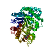
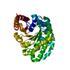
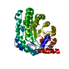
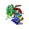
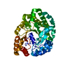
- PDB-6jaw: Crystal structure of Ostrinia furnacalis Group II chitinase catal... -
+ Open data

- Basic information
| Entry | Database: PDB / ID: 6jaw | ||||||
|---|---|---|---|---|---|---|---|
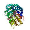
| Title | Crystal structure of Ostrinia furnacalis Group II chitinase catalytic domain 1 in complex with a napthalimide derivative | ||||||
 Components Components | Group II chitinase | ||||||
 Keywords Keywords |  HYDROLASE / inhibitor complex / chitinase HYDROLASE / inhibitor complex / chitinase | ||||||
| Function / homology |  Function and homology informationchitinase / chitinase activity / chitin catabolic process / chitin binding / carbohydrate metabolic process / extracellular region Function and homology informationchitinase / chitinase activity / chitin catabolic process / chitin binding / carbohydrate metabolic process / extracellular regionSimilarity search - Function | ||||||
| Biological species |  Ostrinia furnacalis (Asian corn borer) Ostrinia furnacalis (Asian corn borer) | ||||||
| Method | X-RAY DIFFRACTION / SYNCHROTRON / MOLECULAR REPLACEMENT / Resolution: 1.981 Å | ||||||
 Authors Authors | Chen, W. / Zhou, Y. / Yang, Q. | ||||||
| Funding support |  China, 1items China, 1items
| ||||||
 Citation Citation | Journal: J.Biol.Chem. / Year: 2019 Title: Structural dissection reveals a general mechanistic principle for group II chitinase (ChtII) inhibition. Authors: Chen, W. / Zhou, Y. / Yang, Q. | ||||||
| History |
|
- Structure visualization
| Structure viewer | Molecule: MolmilJmol/JSmol |
|---|
- Downloads & links
-Download
| PDBx/mmCIF format | 6jaw.cif.gz | 98.5 KB | Display | PDBx/mmCIF format |
|---|---|---|---|---|
| PDB format | pdb6jaw.ent.gz | 70.8 KB | Display | PDB format |
| PDBx/mmJSON format | 6jaw.json.gz | Tree view | PDBx/mmJSON format | |
| Others |  Other downloads Other downloads |
-Validation report
| Arichive directory | https://data.pdbj.org/pub/pdb/validation_reports/ja/6jawftp://data.pdbj.org/pub/pdb/validation_reports/ja/6jaw | HTTPS FTP |
|---|
-Related structure data
| Related structure data |  6javC 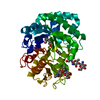 6jaxC 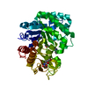 6jayC 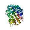 5y29S S: Starting model for refinement C: citing same article ( |
|---|---|
| Similar structure data |


-Links
 PDBj
PDBj- Assembly
| Deposited unit | 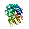
| ||||||||
|---|---|---|---|---|---|---|---|---|---|
| 1 |
| ||||||||
| Unit cell |
|
-Components
| #1: Protein | Mass: 44468.395 Da / Num. of mol.: 1 Source method: isolated from a genetically manipulated source Source: (gene. exp.) Ostrinia furnacalis (Asian corn borer) / Production host:  Komagataella pastoris GS115 (fungus) / References: UniProt: A0A221ZS22, chitinase Komagataella pastoris GS115 (fungus) / References: UniProt: A0A221ZS22, chitinase |
|---|---|
| #2: Chemical | ChemComp-BBO / 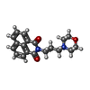  Mass: 324.374 Da / Num. of mol.: 1 / Source method: obtained synthetically / Formula: C19H20N2O3 / Feature type: SUBJECT OF INVESTIGATION Mass: 324.374 Da / Num. of mol.: 1 / Source method: obtained synthetically / Formula: C19H20N2O3 / Feature type: SUBJECT OF INVESTIGATION |
| #3: Sugar | ChemComp-NAG / N-Acetylglucosamine  Type: D-saccharide, beta linking / Mass: 221.208 Da / Num. of mol.: 1 Type: D-saccharide, beta linking / Mass: 221.208 Da / Num. of mol.: 1Source method: isolated from a genetically manipulated source Formula: C8H15NO6 |
| #4: Water | ChemComp-HOH / Water Mass: 18.015 Da / Num. of mol.: 225 / Source method: isolated from a natural source / Formula: H2O Mass: 18.015 Da / Num. of mol.: 225 / Source method: isolated from a natural source / Formula: H2O |
| Sequence details | The residues 1863-1878 (GDKWDSPREQWRKDAN) seriously influenced its expression and crystallization, ...The residues 1863-1878 (GDKWDSPREQ |
-Experimental details
-Experiment
| Experiment | Method: X-RAY DIFFRACTION / Number of used crystals: 1 |
|---|
- Sample preparation
| Crystal | Density Matthews: 2.56 Å3/Da / Density % sol: 51.87 % |
|---|---|
| Crystal grow | Temperature: 277 K / Method: vapor diffusion, hanging drop / pH: 8.5 Details: 0.2M sodium chloride, 0.1M Tris (pH 8.5) and 25% PEG3350 |
-Data collection
| Diffraction | Mean temperature: 100 K / Serial crystal experiment: N |
|---|---|
| Diffraction source | Source: SYNCHROTRON / Site: NFPSS / Beamline: BL18U / Wavelength: 0.9793 Å |
| Detector | Type: DECTRIS PILATUS3 6M / Detector: PIXEL / Date: Jun 2, 2018 |
| Radiation | Protocol: SINGLE WAVELENGTH / Monochromatic (M) / Laue (L): M / Scattering type: x-ray |
| Radiation wavelength | Wavelength: 0.9793 Å / Relative weight: 1 |
| Reflection | Resolution: 1.981→50 Å / Num. obs: 51402 / % possible obs: 100 % / Redundancy: 12.7 % / Rsym value: 0.179 / Net I/σ(I): 4.1 |
| Reflection shell | Resolution: 1.981→2.03 Å / Redundancy: 12.6 % / Num. unique obs: 1595 / Rsym value: 1.318 / % possible all: 100 |
- Processing
| Software |
| ||||||||||||||||||||||||||||||||||||||||||||||||||||||||||||||||||||||||||||||||||||||||||||||||||||||||||||||||||||||||||||||||||||||||||||||||||||||||||||||||||||||||
|---|---|---|---|---|---|---|---|---|---|---|---|---|---|---|---|---|---|---|---|---|---|---|---|---|---|---|---|---|---|---|---|---|---|---|---|---|---|---|---|---|---|---|---|---|---|---|---|---|---|---|---|---|---|---|---|---|---|---|---|---|---|---|---|---|---|---|---|---|---|---|---|---|---|---|---|---|---|---|---|---|---|---|---|---|---|---|---|---|---|---|---|---|---|---|---|---|---|---|---|---|---|---|---|---|---|---|---|---|---|---|---|---|---|---|---|---|---|---|---|---|---|---|---|---|---|---|---|---|---|---|---|---|---|---|---|---|---|---|---|---|---|---|---|---|---|---|---|---|---|---|---|---|---|---|---|---|---|---|---|---|---|---|---|---|---|---|---|---|---|
| Refinement | Method to determine structure: MOLECULAR REPLACEMENT Starting model: 5Y29 Resolution: 1.981→43.566 Å / SU ML: 0.15 / Cross valid method: FREE R-VALUE / σ(F): 1.36 / Phase error: 19.3 Details: SF FILE CONTAINS FRIEDEL PAIRS UNDER I_MINUS AND I_PLUS COLUMNS.
| ||||||||||||||||||||||||||||||||||||||||||||||||||||||||||||||||||||||||||||||||||||||||||||||||||||||||||||||||||||||||||||||||||||||||||||||||||||||||||||||||||||||||
| Solvent computation | Shrinkage radii: 0.9 Å / VDW probe radii: 1.11 Å | ||||||||||||||||||||||||||||||||||||||||||||||||||||||||||||||||||||||||||||||||||||||||||||||||||||||||||||||||||||||||||||||||||||||||||||||||||||||||||||||||||||||||
| Refinement step | Cycle: LAST / Resolution: 1.981→43.566 Å
| ||||||||||||||||||||||||||||||||||||||||||||||||||||||||||||||||||||||||||||||||||||||||||||||||||||||||||||||||||||||||||||||||||||||||||||||||||||||||||||||||||||||||
| Refine LS restraints |
| ||||||||||||||||||||||||||||||||||||||||||||||||||||||||||||||||||||||||||||||||||||||||||||||||||||||||||||||||||||||||||||||||||||||||||||||||||||||||||||||||||||||||
| LS refinement shell |
|
