 Movie
Movie Controller


+ Open data

- Basic information
| Entry | Database: PDB / ID: 6iih | ||||||
|---|---|---|---|---|---|---|---|
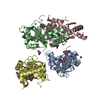
| Title | crystal structure of mitochondrial calcium uptake 2(MICU2) | ||||||
 Components Components | Endolysin,Calcium uptake protein 2, mitochondrial | ||||||
 Keywords Keywords |  Calcium Binding Protein / mitochondrial / EF-hand / T4L fusion protein / METAL BINDING PROTEIN Calcium Binding Protein / mitochondrial / EF-hand / T4L fusion protein / METAL BINDING PROTEIN | ||||||
| Function / homology |  Function and homology information Function and homology informationnegative regulation of mitochondrial calcium ion concentration / Processing of SMDT1 / mitochondrial calcium ion transmembrane transport / uniplex complex / calcium import into the mitochondrion / Mitochondrial calcium ion transport / positive regulation of mitochondrial calcium ion concentration / mitochondrial calcium ion homeostasis / viral release from host cell by cytolysis / calcium channel complex ...negative regulation of mitochondrial calcium ion concentration / Processing of SMDT1 / mitochondrial calcium ion transmembrane transport / uniplex complex / calcium import into the mitochondrion / Mitochondrial calcium ion transport / positive regulation of mitochondrial calcium ion concentration / mitochondrial calcium ion homeostasis / viral release from host cell by cytolysis / calcium channel complex / peptidoglycan catabolic process / mitochondrial intermembrane space / cell wall macromolecule catabolic process / lysozyme / lysozyme activity / mitochondrial inner membrane / host cell cytoplasm / defense response to bacterium / protein heterodimerization activity / calcium ion binding / mitochondrionSimilarity search - Function | ||||||
| Biological species |  Enterobacteria phage T4 (virus) Enterobacteria phage T4 (virus) Homo sapiens (human) Homo sapiens (human) | ||||||
| Method | X-RAY DIFFRACTION / SYNCHROTRON / MOLECULAR REPLACEMENT / Resolution: 1.958 Å | ||||||
 Authors Authors | Shen, Q. / Wu, W. / Zheng, J. / Jia, Z. | ||||||
| Funding support |  China, 1items China, 1items
| ||||||
 Citation Citation | Journal: Embo Rep. / Year: 2019 Title: The crystal structure of MICU2 provides insight into Ca2+binding and MICU1-MICU2 heterodimer formation. Authors: Wu, W. / Shen, Q. / Lei, Z. / Qiu, Z. / Li, D. / Pei, H. / Zheng, J. / Jia, Z. | ||||||
| History |
|
- Structure visualization
| Structure viewer | Molecule: MolmilJmol/JSmol |
|---|
- Downloads & links
-Download
| PDBx/mmCIF format | 6iih.cif.gz | 368.9 KB | Display | PDBx/mmCIF format |
|---|---|---|---|---|
| PDB format | pdb6iih.ent.gz | 300.6 KB | Display | PDB format |
| PDBx/mmJSON format | 6iih.json.gz | Tree view | PDBx/mmJSON format | |
| Others |  Other downloads Other downloads |
-Validation report
| Arichive directory | https://data.pdbj.org/pub/pdb/validation_reports/ii/6iihftp://data.pdbj.org/pub/pdb/validation_reports/ii/6iih | HTTPS FTP |
|---|
-Related structure data
| Similar structure data |
|---|


-Links
 PDBj
PDBj


- Assembly
| Deposited unit | 
| ||||||||
|---|---|---|---|---|---|---|---|---|---|
| 1 |
| ||||||||
| Unit cell |
|
-Components
| #1: Protein | Mass: 57739.125 Da / Num. of mol.: 2 / Mutation: C54T, C97A Source method: isolated from a genetically manipulated source Source: (gene. exp.) Enterobacteria phage T4 (virus), (gene. exp.) Homo sapiens (human)Plasmid: pET-28(b) / Gene: MICU2, EFHA1 / Production host:  Escherichia coli BL21(DE3) (bacteria) / References: UniProt: P00720, UniProt: Q8IYU8, lysozyme Escherichia coli BL21(DE3) (bacteria) / References: UniProt: P00720, UniProt: Q8IYU8, lysozyme#2: Chemical |   Mass: 40.078 Da / Num. of mol.: 3 / Source method: obtained synthetically / Formula: Ca Mass: 40.078 Da / Num. of mol.: 3 / Source method: obtained synthetically / Formula: Ca#3: Water | ChemComp-HOH / | Water Mass: 18.015 Da / Num. of mol.: 819 / Source method: isolated from a natural source / Formula: H2O Mass: 18.015 Da / Num. of mol.: 819 / Source method: isolated from a natural source / Formula: H2O |
|---|
-Experimental details
-Experiment
| Experiment | Method: X-RAY DIFFRACTION / Number of used crystals: 1 |
|---|
- Sample preparation
| Crystal | Density Matthews: 2.39 Å3/Da / Density % sol: 48.59 % |
|---|---|
| Crystal grow | Temperature: 293 K / Method: vapor diffusion, sitting drop / pH: 5.16 Details: 100 mM Sodium phosphate monobasic , 100 mM potassium phosphate monobasic , 100 mM MES, 6.0 and 1M sodium chloride |
-Data collection
| Diffraction | Mean temperature: 100 K / Serial crystal experiment: N |
|---|---|
| Diffraction source | Source: SYNCHROTRON / Site: SSRF / Beamline: BL19U1 / Wavelength: 0.978 Å |
| Detector | Type: DECTRIS PILATUS3 6M / Detector: PIXEL / Date: Dec 24, 2017 |
| Radiation | Protocol: SINGLE WAVELENGTH / Monochromatic (M) / Laue (L): M / Scattering type: x-ray |
| Radiation wavelength | Wavelength: 0.978 Å / Relative weight: 1 |
| Reflection | Resolution: 1.958→48.676 Å / Num. obs: 73695 / % possible obs: 96 % / Redundancy: 2.8 % / CC1/2: 0.997 / Rmerge(I) obs: 0.048 / Rpim(I) all: 0.033 / Rrim(I) all: 0.058 / Χ2: 0.977 / Net I/av σ(I): 21.26 / Net I/σ(I): 8.98 |
| Reflection shell | Resolution: 1.958→2.007 Å / Redundancy: 2.8 % / Rmerge(I) obs: 0.507 / Mean I/σ(I) obs: 1.65 / Num. unique obs: 3701 / CC1/2: 0.692 / Rpim(I) all: 0.365 / Rrim(I) all: 0.629 / Χ2: 0.971 / % possible all: 97.9 |
- Processing
| Software |
| |||||||||||||||||||||||||||||||||||||||||||||||||||||||||||||||||||||||||||||||||||||||||||||||||||||||||
|---|---|---|---|---|---|---|---|---|---|---|---|---|---|---|---|---|---|---|---|---|---|---|---|---|---|---|---|---|---|---|---|---|---|---|---|---|---|---|---|---|---|---|---|---|---|---|---|---|---|---|---|---|---|---|---|---|---|---|---|---|---|---|---|---|---|---|---|---|---|---|---|---|---|---|---|---|---|---|---|---|---|---|---|---|---|---|---|---|---|---|---|---|---|---|---|---|---|---|---|---|---|---|---|---|---|---|
| Refinement | Method to determine structure: MOLECULAR REPLACEMENT / Resolution: 1.958→48.676 Å / SU ML: 0.22 / Cross valid method: FREE R-VALUE / σ(F): 1.35 / Phase error: 22.17 / Stereochemistry target values: ML
| |||||||||||||||||||||||||||||||||||||||||||||||||||||||||||||||||||||||||||||||||||||||||||||||||||||||||
| Solvent computation | Shrinkage radii: 0.9 Å / VDW probe radii: 1.11 Å / Solvent model: FLAT BULK SOLVENT MODEL | |||||||||||||||||||||||||||||||||||||||||||||||||||||||||||||||||||||||||||||||||||||||||||||||||||||||||
| Refinement step | Cycle: LAST / Resolution: 1.958→48.676 Å
| |||||||||||||||||||||||||||||||||||||||||||||||||||||||||||||||||||||||||||||||||||||||||||||||||||||||||
| Refine LS restraints |
| |||||||||||||||||||||||||||||||||||||||||||||||||||||||||||||||||||||||||||||||||||||||||||||||||||||||||
| LS refinement shell |
|
