 Movie
Movie Controller

[English] 日本語
 Yorodumi
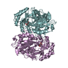
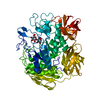
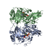
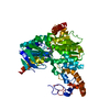
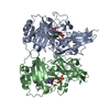
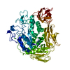
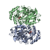
- PDB-6ig2: Structure of mitochondrial CDP-DAG synthase Tam41 complexed with ... -
+ Open data

- Basic information
| Entry | Database: PDB / ID: 6ig2 | |||||||||
|---|---|---|---|---|---|---|---|---|---|---|
| Title | Structure of mitochondrial CDP-DAG synthase Tam41 complexed with CTP, delta 74, F240A | |||||||||
 Components Components | Phosphatidate cytidylyltransferase, mitochondrial | |||||||||
 Keywords Keywords | TRANSFERASE / mitochondrial inner membrane / CDP-Diacylglycerol synthase / NTase fold | |||||||||
| Function / homology |  Function and homology informationphosphatidate cytidylyltransferase / phosphatidate cytidylyltransferase activity / extrinsic component of mitochondrial inner membrane / CDP-diacylglycerol biosynthetic process / cardiolipin biosynthetic process / cardiolipin binding / phosphatidic acid binding / mitochondrial membrane organization / mitochondrial matrix / mitochondrion Function and homology informationphosphatidate cytidylyltransferase / phosphatidate cytidylyltransferase activity / extrinsic component of mitochondrial inner membrane / CDP-diacylglycerol biosynthetic process / cardiolipin biosynthetic process / cardiolipin binding / phosphatidic acid binding / mitochondrial membrane organization / mitochondrial matrix / mitochondrionSimilarity search - Function | |||||||||
| Biological species |  Schizosaccharomyces pombe (fission yeast) Schizosaccharomyces pombe (fission yeast) | |||||||||
| Method | X-RAY DIFFRACTION / SYNCHROTRON / MOLECULAR REPLACEMENT / Resolution: 2.882 Å | |||||||||
 Authors Authors | Jiao, H.Z. / Yin, Y. / Liu, Z.F. | |||||||||
| Funding support |  China, 2items China, 2items
| |||||||||
 Citation Citation | Journal: Structure / Year: 2019 Title: Structures of the Mitochondrial CDP-DAG Synthase Tam41 Suggest a Potential Lipid Substrate Pathway from Membrane to the Active Site. Authors: Jiao, H. / Yin, Y. / Liu, Z. | |||||||||
| History |
|
- Structure visualization
| Structure viewer | Molecule: MolmilJmol/JSmol |
|---|
- Downloads & links
-Download
| PDBx/mmCIF format | 6ig2.cif.gz | 237.6 KB | Display | PDBx/mmCIF format |
|---|---|---|---|---|
| PDB format | pdb6ig2.ent.gz | 189.5 KB | Display | PDB format |
| PDBx/mmJSON format | 6ig2.json.gz | Tree view | PDBx/mmJSON format | |
| Others |  Other downloads Other downloads |
-Validation report
| Arichive directory | https://data.pdbj.org/pub/pdb/validation_reports/ig/6ig2ftp://data.pdbj.org/pub/pdb/validation_reports/ig/6ig2 | HTTPS FTP |
|---|
-Related structure data
| Related structure data | 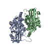 6ig4SC S: Starting model for refinement C: citing same article ( |
|---|---|
| Similar structure data |


-Links
 PDBj
PDBj
- Assembly
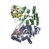
| Deposited unit | 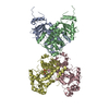
| ||||||||
|---|---|---|---|---|---|---|---|---|---|
| 1 | 
| ||||||||
| 2 | 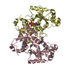
| ||||||||
| Unit cell |
|
-Components
| #1: Protein | / Tam41 / CDP-diacylglycerol synthase / CDP-DAG synthase / Mitochondrial translocator assembly and ...Tam41 / CDP-diacylglycerol synthase / CDP-DAG synthase / Mitochondrial translocator assembly and maintenance protein 41 homolog Mass: 35856.719 Da / Num. of mol.: 4 / Mutation: F240A Source method: isolated from a genetically manipulated source Source: (gene. exp.) Schizosaccharomyces pombe (strain 972 / ATCC 24843) (yeast)Strain: 972 / ATCC 24843 / Gene: tam41, SPBC1A4.06c / Production host:  Escherichia coli (E. coli) Escherichia coli (E. coli)References: UniProt: O74339, phosphatidate cytidylyltransferase#2: Chemical | ChemComp-CTP / | Cytidine triphosphate  Mass: 483.156 Da / Num. of mol.: 1 / Source method: obtained synthetically / Formula: C9H16N3O14P3 Mass: 483.156 Da / Num. of mol.: 1 / Source method: obtained synthetically / Formula: C9H16N3O14P3#3: Water | ChemComp-HOH / | Water Mass: 18.015 Da / Num. of mol.: 216 / Source method: isolated from a natural source / Formula: H2O Mass: 18.015 Da / Num. of mol.: 216 / Source method: isolated from a natural source / Formula: H2O |
|---|
-Experimental details
-Experiment
| Experiment | Method: X-RAY DIFFRACTION / Number of used crystals: 1 |
|---|
- Sample preparation
| Crystal | Density Matthews: 3.37 Å3/Da / Density % sol: 63.53 % |
|---|---|
| Crystal grow | Temperature: 289.15 K / Method: evaporation, recrystallization Details: 0.1 M HEPES-Na, pH 7.5, 9% Tacsimate, pH 7.0, 2% PEG 3350, incubated with 10 mM MgCl2, 5 mM CTP for 1 h before crystallization PH range: 7.0-7.5 |
-Data collection
| Diffraction | Mean temperature: 173.15 K / Serial crystal experiment: N |
|---|---|
| Diffraction source | Source: SYNCHROTRON / Site: SSRF / Beamline: BL18U1 / Wavelength: 0.977 Å |
| Detector | Type: DECTRIS PILATUS3 6M / Detector: PIXEL / Date: Jun 24, 2017 |
| Radiation | Protocol: SINGLE WAVELENGTH / Monochromatic (M) / Laue (L): M / Scattering type: x-ray |
| Radiation wavelength | Wavelength: 0.977 Å / Relative weight: 1 |
| Reflection | Resolution: 2.88→50 Å / Num. obs: 44684 / % possible obs: 100 % / Redundancy: 6.5 % / Rpim(I) all: 0.042 / Net I/σ(I): 20.7 |
| Reflection shell | Resolution: 2.9→3 Å / Redundancy: 6.8 % / Mean I/σ(I) obs: 1.9 / Num. unique obs: 4338 / Rpim(I) all: 0.482 / % possible all: 100 |
- Processing
| Software |
| ||||||||||||||||||||||||||||||||||||||||||||||||||||||||||||||||||||||||||||||||||||||||||||||||||||||||||||||||
|---|---|---|---|---|---|---|---|---|---|---|---|---|---|---|---|---|---|---|---|---|---|---|---|---|---|---|---|---|---|---|---|---|---|---|---|---|---|---|---|---|---|---|---|---|---|---|---|---|---|---|---|---|---|---|---|---|---|---|---|---|---|---|---|---|---|---|---|---|---|---|---|---|---|---|---|---|---|---|---|---|---|---|---|---|---|---|---|---|---|---|---|---|---|---|---|---|---|---|---|---|---|---|---|---|---|---|---|---|---|---|---|---|---|
| Refinement | Method to determine structure: MOLECULAR REPLACEMENT Starting model: 6IG4 Resolution: 2.882→47.604 Å / SU ML: 0.33 / Cross valid method: FREE R-VALUE / σ(F): 1.34 / Phase error: 25.92
| ||||||||||||||||||||||||||||||||||||||||||||||||||||||||||||||||||||||||||||||||||||||||||||||||||||||||||||||||
| Solvent computation | Shrinkage radii: 0.9 Å / VDW probe radii: 1.11 Å | ||||||||||||||||||||||||||||||||||||||||||||||||||||||||||||||||||||||||||||||||||||||||||||||||||||||||||||||||
| Refinement step | Cycle: LAST / Resolution: 2.882→47.604 Å
| ||||||||||||||||||||||||||||||||||||||||||||||||||||||||||||||||||||||||||||||||||||||||||||||||||||||||||||||||
| Refine LS restraints |
| ||||||||||||||||||||||||||||||||||||||||||||||||||||||||||||||||||||||||||||||||||||||||||||||||||||||||||||||||
| LS refinement shell |
|
