 Movie
Movie Controller

[English] 日本語
 Yorodumi
- PDB-6hv6: Crystal structure of PatoxP, a cysteine protease-like domain of P... -
+ Open data

- Basic information
| Entry | Database: PDB / ID: 6hv6 | |||||||||
|---|---|---|---|---|---|---|---|---|---|---|
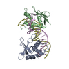
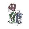
| Title | Crystal structure of PatoxP, a cysteine protease-like domain of Photorhabdus asymbiotica toxin PaTox | |||||||||
 Components Components | Toxin PAU_02230 | |||||||||
 Keywords Keywords |  TOXIN / Cystein protease-like / PaTox toxin TOXIN / Cystein protease-like / PaTox toxin | |||||||||
| Function / homology |  Function and homology information Function and homology information: / protein N-acetylglucosaminyltransferase activity / protein-glutamine glutaminase activity / protein-glutamine glutaminase / Transferases; Glycosyltransferases; Hexosyltransferases / regulation of GTPase activity / metabolic process / toxin activity / calcium ion binding / host cell plasma membrane ...: / protein N-acetylglucosaminyltransferase activity / protein-glutamine glutaminase activity / protein-glutamine glutaminase / Transferases; Glycosyltransferases; Hexosyltransferases / regulation of GTPase activity / metabolic process / toxin activity / calcium ion binding / host cell plasma membrane / extracellular region / membraneSimilarity search - Function | |||||||||
| Biological species |  Photorhabdus asymbiotica subsp. asymbiotica ATCC 43949 (bacteria) Photorhabdus asymbiotica subsp. asymbiotica ATCC 43949 (bacteria) | |||||||||
| Method | X-RAY DIFFRACTION / SYNCHROTRON / SAD / Resolution: 2.001 Å | |||||||||
 Authors Authors | Bogdanovic, X. / Wirth, C. / Hunte, C. | |||||||||
| Funding support |  Germany, 2items Germany, 2items
| |||||||||
 Citation Citation | Journal: J. Biol. Chem. / Year: 2019 Title: A cysteine protease-like domain enhances the cytotoxic effects of thePhotorhabdus asymbioticatoxin PaTox. Authors: Bogdanovic, X. / Schneider, S. / Levanova, N. / Wirth, C. / Trillhaase, C. / Steinemann, M. / Hunte, C. / Aktories, K. / Jank, T. | |||||||||
| History |
|
- Structure visualization
| Structure viewer | Molecule: MolmilJmol/JSmol |
|---|
- Downloads & links
-Download
| PDBx/mmCIF format | 6hv6.cif.gz | 138.3 KB | Display | PDBx/mmCIF format |
|---|---|---|---|---|
| PDB format | pdb6hv6.ent.gz | 108.5 KB | Display | PDB format |
| PDBx/mmJSON format | 6hv6.json.gz | Tree view | PDBx/mmJSON format | |
| Others |  Other downloads Other downloads |
-Validation report
| Arichive directory | https://data.pdbj.org/pub/pdb/validation_reports/hv/6hv6ftp://data.pdbj.org/pub/pdb/validation_reports/hv/6hv6 | HTTPS FTP |
|---|
-Related structure data
| Similar structure data |
|---|


-Links
 PDBj
PDBj


- Assembly
| Deposited unit | 
| ||||||||
|---|---|---|---|---|---|---|---|---|---|
| 1 |
| ||||||||
| Unit cell |
|
-Components
| #1: Protein | Mass: 52282.512 Da / Num. of mol.: 1 / Mutation: C1865A Source method: isolated from a genetically manipulated source Details: The catalytic cysteine 1865 was mutated to alanine Source: (gene. exp.) Photorhabdus asymbiotica subsp. asymbiotica ATCC 43949 (bacteria)Gene: PAU_02230 / Production host: Escherichia coli BL21(DE3) (bacteria)References: UniProt: C7BKP9, Transferases; Glycosyltransferases; Hexosyltransferases, protein-glutamine glutaminase | ||||
|---|---|---|---|---|---|
| #2: Chemical | ChemComp-EDO / Ethylene glycol  Mass: 62.068 Da / Num. of mol.: 6 / Source method: obtained synthetically / Formula: C2H6O2 Mass: 62.068 Da / Num. of mol.: 6 / Source method: obtained synthetically / Formula: C2H6O2#3: Chemical | ChemComp-EPE / | HEPES  Mass: 238.305 Da / Num. of mol.: 1 / Source method: obtained synthetically / Formula: C8H18N2O4S / Comment: pH buffer*YM Mass: 238.305 Da / Num. of mol.: 1 / Source method: obtained synthetically / Formula: C8H18N2O4S / Comment: pH buffer*YM#4: Water | ChemComp-HOH / | Water Mass: 18.015 Da / Num. of mol.: 184 / Source method: isolated from a natural source / Formula: H2O Mass: 18.015 Da / Num. of mol.: 184 / Source method: isolated from a natural source / Formula: H2O |
-Experimental details
-Experiment
| Experiment | Method: X-RAY DIFFRACTION / Number of used crystals: 1 |
|---|
- Sample preparation
| Crystal grow | Temperature: 293 K / Method: vapor diffusion, sitting drop / pH: 7.4 Details: 25.7% w/v Polyethylene glycol 8000, 150 mM HEPES pH 7.5, 60 mM potassium sodium tartrate |
|---|
-Data collection
| Diffraction | Mean temperature: 100 K / Serial crystal experiment: N |
|---|---|
| Diffraction source | Source: SYNCHROTRON / Site: SLS  / Beamline: X06SA / Wavelength: 1.2037 Å / Beamline: X06SA / Wavelength: 1.2037 Å |
| Detector | Type: DECTRIS EIGER X 16M / Detector: PIXEL / Date: Nov 21, 2016 |
| Radiation | Protocol: SINGLE WAVELENGTH / Monochromatic (M) / Laue (L): M / Scattering type: x-ray |
| Radiation wavelength | Wavelength: 1.2037 Å / Relative weight: 1 |
| Reflection | Resolution: 2→53.9 Å / Num. obs: 25589 / % possible obs: 100 % / Redundancy: 95.5 % / CC1/2: 0.997 / Rmerge(I) obs: 0.261 / Net I/σ(I): 14.7 |
| Reflection shell | Resolution: 2→2.05 Å / Redundancy: 62 % / Rmerge(I) obs: 2.298 / Mean I/σ(I) obs: 2.5 / Num. unique obs: 1856 / CC1/2: 0.772 / % possible all: 99.9 |
- Processing
| Software |
| ||||||||||||||||||||||||||||||||||||||||||||||||||||||||||||||||||||||||||||||||||||||||||||||||||||||||||||||||||||||||||||||||||||||||||||||||||||||||||||||||||||||||||||||||||||||||||||||||||||||||
|---|---|---|---|---|---|---|---|---|---|---|---|---|---|---|---|---|---|---|---|---|---|---|---|---|---|---|---|---|---|---|---|---|---|---|---|---|---|---|---|---|---|---|---|---|---|---|---|---|---|---|---|---|---|---|---|---|---|---|---|---|---|---|---|---|---|---|---|---|---|---|---|---|---|---|---|---|---|---|---|---|---|---|---|---|---|---|---|---|---|---|---|---|---|---|---|---|---|---|---|---|---|---|---|---|---|---|---|---|---|---|---|---|---|---|---|---|---|---|---|---|---|---|---|---|---|---|---|---|---|---|---|---|---|---|---|---|---|---|---|---|---|---|---|---|---|---|---|---|---|---|---|---|---|---|---|---|---|---|---|---|---|---|---|---|---|---|---|---|---|---|---|---|---|---|---|---|---|---|---|---|---|---|---|---|---|---|---|---|---|---|---|---|---|---|---|---|---|---|---|---|---|
| Refinement | Method to determine structure: SAD / Resolution: 2.001→53.864 Å / SU ML: 0.2 / Cross valid method: FREE R-VALUE / σ(F): 1.34 / Phase error: 20.82
| ||||||||||||||||||||||||||||||||||||||||||||||||||||||||||||||||||||||||||||||||||||||||||||||||||||||||||||||||||||||||||||||||||||||||||||||||||||||||||||||||||||||||||||||||||||||||||||||||||||||||
| Solvent computation | Shrinkage radii: 0.9 Å / VDW probe radii: 1.11 Å | ||||||||||||||||||||||||||||||||||||||||||||||||||||||||||||||||||||||||||||||||||||||||||||||||||||||||||||||||||||||||||||||||||||||||||||||||||||||||||||||||||||||||||||||||||||||||||||||||||||||||
| Refinement step | Cycle: LAST / Resolution: 2.001→53.864 Å
| ||||||||||||||||||||||||||||||||||||||||||||||||||||||||||||||||||||||||||||||||||||||||||||||||||||||||||||||||||||||||||||||||||||||||||||||||||||||||||||||||||||||||||||||||||||||||||||||||||||||||
| Refine LS restraints |
| ||||||||||||||||||||||||||||||||||||||||||||||||||||||||||||||||||||||||||||||||||||||||||||||||||||||||||||||||||||||||||||||||||||||||||||||||||||||||||||||||||||||||||||||||||||||||||||||||||||||||
| LS refinement shell |
| ||||||||||||||||||||||||||||||||||||||||||||||||||||||||||||||||||||||||||||||||||||||||||||||||||||||||||||||||||||||||||||||||||||||||||||||||||||||||||||||||||||||||||||||||||||||||||||||||||||||||
| Refinement TLS params. | Method: refined / Refine-ID: X-RAY DIFFRACTION
| ||||||||||||||||||||||||||||||||||||||||||||||||||||||||||||||||||||||||||||||||||||||||||||||||||||||||||||||||||||||||||||||||||||||||||||||||||||||||||||||||||||||||||||||||||||||||||||||||||||||||
| Refinement TLS group |
|
