 Movie
Movie Controller

[English] 日本語
 Yorodumi
- PDB-6ht1: Crystal structure of MLLT1 (ENL) YEATS domain in complexed with S... -
+ Open data

- Basic information
| Entry | Database: PDB / ID: 6ht1 | ||||||
|---|---|---|---|---|---|---|---|
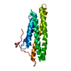
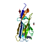
| Title | Crystal structure of MLLT1 (ENL) YEATS domain in complexed with SGC-iMLLT (compound 92) | ||||||
 Components Components | Protein ENL | ||||||
 Keywords Keywords |  TRANSCRIPTION / YEATS domain / ENL / MLLT1 / chemical probe / inhibitor / Structural Genomics / Structural Genomics Consortium / SGC TRANSCRIPTION / YEATS domain / ENL / MLLT1 / chemical probe / inhibitor / Structural Genomics / Structural Genomics Consortium / SGC | ||||||
| Function / homology |  Function and homology information Function and homology informationRNA Polymerase II Transcription Elongation / Formation of RNA Pol II elongation complex / RNA Polymerase II Pre-transcription Events / transcription elongation factor complex / lysine-acetylated histone binding / fibrillar center / chromatin binding / positive regulation of DNA-templated transcription / nucleoplasm / cytosolSimilarity search - Function | ||||||
| Biological species |  Homo sapiens (human) Homo sapiens (human) | ||||||
| Method | X-RAY DIFFRACTION / SYNCHROTRON / MOLECULAR REPLACEMENT / Resolution: 2.1 Å | ||||||
 Authors Authors | Heidenreich, D. / Chaikuad, A. / Moustakim, M. / Arrowsmith, C.H. / Edwards, A.M. / Bountra, C. / Fedorov, O. / Brennan, P.E. / Knapp, S. / Structural Genomics Consortium (SGC) | ||||||
 Citation Citation | Journal: Angew. Chem. Int. Ed. Engl. / Year: 2018 Title: Discovery of an MLLT1/3 YEATS Domain Chemical Probe. Authors: Moustakim, M. / Christott, T. / Monteiro, O.P. / Bennett, J. / Giroud, C. / Ward, J. / Rogers, C.M. / Smith, P. / Panagakou, I. / Diaz-Saez, L. / Felce, S.L. / Gamble, V. / Gileadi, C. / ...Authors: Moustakim, M. / Christott, T. / Monteiro, O.P. / Bennett, J. / Giroud, C. / Ward, J. / Rogers, C.M. / Smith, P. / Panagakou, I. / Diaz-Saez, L. / Felce, S.L. / Gamble, V. / Gileadi, C. / Halidi, N. / Heidenreich, D. / Chaikuad, A. / Knapp, S. / Huber, K.V.M. / Farnie, G. / Heer, J. / Manevski, N. / Poda, G. / Al-Awar, R. / Dixon, D.J. / Brennan, P.E. / Fedorov, O. | ||||||
| History |
|
- Structure visualization
| Structure viewer | Molecule: MolmilJmol/JSmol |
|---|
- Downloads & links
-Download
| PDBx/mmCIF format | 6ht1.cif.gz | 80.1 KB | Display | PDBx/mmCIF format |
|---|---|---|---|---|
| PDB format | pdb6ht1.ent.gz | 59.5 KB | Display | PDB format |
| PDBx/mmJSON format | 6ht1.json.gz | Tree view | PDBx/mmJSON format | |
| Others |  Other downloads Other downloads |
-Validation report
| Arichive directory | https://data.pdbj.org/pub/pdb/validation_reports/ht/6ht1ftp://data.pdbj.org/pub/pdb/validation_reports/ht/6ht1 | HTTPS FTP |
|---|
-Related structure data
| Related structure data |  6ht0C  5j9sS C: citing same article ( S: Starting model for refinement |
|---|---|
| Similar structure data |

-Links
 PDBj
PDBj

- Assembly
| Deposited unit | 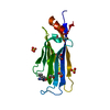
| ||||||||
|---|---|---|---|---|---|---|---|---|---|
| 1 |
| ||||||||
| Unit cell |
|
-Components
| #1: Protein | Mass: 18312.080 Da / Num. of mol.: 1 Source method: isolated from a genetically manipulated source Source: (gene. exp.) Homo sapiens (human) / Gene: MLLT1, ENL, LTG19, YEATS1 / Plasmid: pNIC-CH / Production host:  Escherichia coli (E. coli) / Strain (production host): BL21(DE3) / Variant (production host): Rosetta / References: UniProt: Q03111 Escherichia coli (E. coli) / Strain (production host): BL21(DE3) / Variant (production host): Rosetta / References: UniProt: Q03111 | ||||||
|---|---|---|---|---|---|---|---|
| #2: Chemical | Sulfate  Mass: 96.063 Da / Num. of mol.: 2 / Source method: obtained synthetically / Formula: SO4 Mass: 96.063 Da / Num. of mol.: 2 / Source method: obtained synthetically / Formula: SO4#3: Chemical | ChemComp-EDO / Ethylene glycol  Mass: 62.068 Da / Num. of mol.: 7 / Source method: obtained synthetically / Formula: C2H6O2 Mass: 62.068 Da / Num. of mol.: 7 / Source method: obtained synthetically / Formula: C2H6O2#4: Chemical | ChemComp-GQ5 / |   Mass: 388.466 Da / Num. of mol.: 1 / Source method: obtained synthetically / Formula: C22H24N6O Mass: 388.466 Da / Num. of mol.: 1 / Source method: obtained synthetically / Formula: C22H24N6O#5: Water | ChemComp-HOH / | Water Mass: 18.015 Da / Num. of mol.: 51 / Source method: isolated from a natural source / Formula: H2O Mass: 18.015 Da / Num. of mol.: 51 / Source method: isolated from a natural source / Formula: H2O |
-Experimental details
-Experiment
| Experiment | Method: X-RAY DIFFRACTION / Number of used crystals: 1 |
|---|
- Sample preparation
| Crystal | Density Matthews: 2.17 Å3/Da / Density % sol: 43.41 % |
|---|---|
| Crystal grow | Temperature: 293.15 K / Method: vapor diffusion, sitting drop Details: 25% PEG3350, 0.2M ammonium sulfate, 0.1M bis-tris pH 5.5 or 25% PEG Smear Medi-um, 0.1M citrate pH 5.5 |
-Data collection
| Diffraction | Mean temperature: 100 K / Serial crystal experiment: N |
|---|---|
| Diffraction source | Source: SYNCHROTRON / Site: SLS  / Beamline: X06SA / Wavelength: 1 Å / Beamline: X06SA / Wavelength: 1 Å |
| Detector | Type: DECTRIS EIGER X 16M / Detector: PIXEL / Date: Sep 2, 2018 |
| Radiation | Protocol: SINGLE WAVELENGTH / Monochromatic (M) / Laue (L): M / Scattering type: x-ray |
| Radiation wavelength | Wavelength: 1 Å / Relative weight: 1 |
| Reflection | Resolution: 2.1→48.84 Å / Num. obs: 10098 / % possible obs: 100 % / Redundancy: 7.8 % / Biso Wilson estimate: 37.3 Å2 / CC1/2: 0.999 / Rmerge(I) obs: 0.069 / Rpim(I) all: 0.027 / Net I/σ(I): 13.2 |
| Reflection shell | Resolution: 2.1→2.17 Å / Redundancy: 7.6 % / Rmerge(I) obs: 0.463 / Mean I/σ(I) obs: 3.4 / Num. unique obs: 956 / CC1/2: 0.959 / Rpim(I) all: 0.185 / % possible all: 100 |
- Processing
| Software |
| ||||||||||||||||||||||||||||||||||||||||||||||||||||||||||||||||||||||||||||||||||||||||||||||||||||||||||||||||||||||||||||||||||||||||||||||||||||||||||||||||||||||||||||||||||||||
|---|---|---|---|---|---|---|---|---|---|---|---|---|---|---|---|---|---|---|---|---|---|---|---|---|---|---|---|---|---|---|---|---|---|---|---|---|---|---|---|---|---|---|---|---|---|---|---|---|---|---|---|---|---|---|---|---|---|---|---|---|---|---|---|---|---|---|---|---|---|---|---|---|---|---|---|---|---|---|---|---|---|---|---|---|---|---|---|---|---|---|---|---|---|---|---|---|---|---|---|---|---|---|---|---|---|---|---|---|---|---|---|---|---|---|---|---|---|---|---|---|---|---|---|---|---|---|---|---|---|---|---|---|---|---|---|---|---|---|---|---|---|---|---|---|---|---|---|---|---|---|---|---|---|---|---|---|---|---|---|---|---|---|---|---|---|---|---|---|---|---|---|---|---|---|---|---|---|---|---|---|---|---|---|
| Refinement | Method to determine structure: MOLECULAR REPLACEMENT Starting model: 5J9S Resolution: 2.1→45.86 Å / Cor.coef. Fo:Fc: 0.958 / Cor.coef. Fo:Fc free: 0.93 / SU B: 15.601 / SU ML: 0.195 / Cross valid method: THROUGHOUT / ESU R: 0.237 / ESU R Free: 0.2 / Details: HYDROGENS HAVE BEEN ADDED IN THE RIDING POSITIONS
| ||||||||||||||||||||||||||||||||||||||||||||||||||||||||||||||||||||||||||||||||||||||||||||||||||||||||||||||||||||||||||||||||||||||||||||||||||||||||||||||||||||||||||||||||||||||
| Solvent computation | Ion probe radii: 0.8 Å / Shrinkage radii: 0.8 Å / VDW probe radii: 1.2 Å | ||||||||||||||||||||||||||||||||||||||||||||||||||||||||||||||||||||||||||||||||||||||||||||||||||||||||||||||||||||||||||||||||||||||||||||||||||||||||||||||||||||||||||||||||||||||
| Displacement parameters | Biso mean: 53.746 Å2
| ||||||||||||||||||||||||||||||||||||||||||||||||||||||||||||||||||||||||||||||||||||||||||||||||||||||||||||||||||||||||||||||||||||||||||||||||||||||||||||||||||||||||||||||||||||||
| Refinement step | Cycle: 1 / Resolution: 2.1→45.86 Å
| ||||||||||||||||||||||||||||||||||||||||||||||||||||||||||||||||||||||||||||||||||||||||||||||||||||||||||||||||||||||||||||||||||||||||||||||||||||||||||||||||||||||||||||||||||||||
| Refine LS restraints |
|