 Movie
Movie Controller

[English] 日本語
 Yorodumi
- PDB-6hks: Crystal structure of the PTPN3 PDZ domain bound to the HPV16 E6 o... -
+ Open data

- Basic information
| Entry | Database: PDB / ID: 6hks | ||||||
|---|---|---|---|---|---|---|---|
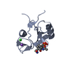
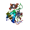
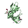
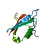
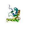
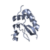
| Title | Crystal structure of the PTPN3 PDZ domain bound to the HPV16 E6 oncoprotein C-terminal peptide | ||||||
 Components Components |
| ||||||
 Keywords Keywords |  HYDROLASE / PDZ domain Protein tyrosine phosphatase Human papillomavirus E6 oncoprotein PDZ binding motif HYDROLASE / PDZ domain Protein tyrosine phosphatase Human papillomavirus E6 oncoprotein PDZ binding motif | ||||||
| Function / homology |  Function and homology information Function and homology informationregulation of membrane depolarization during action potential / symbiont-mediated suppression of host transcription / negative regulation of membrane protein ectodomain proteolysis / regulation of sodium ion transmembrane transporter activity / regulation of proteolysis / activation of GTPase activity / negative regulation of epidermal growth factor receptor signaling pathway / negative regulation of mitotic cell cycle / sodium channel regulator activity / cytoskeletal protein binding ...regulation of membrane depolarization during action potential / symbiont-mediated suppression of host transcription / negative regulation of membrane protein ectodomain proteolysis / regulation of sodium ion transmembrane transporter activity / regulation of proteolysis / activation of GTPase activity / negative regulation of epidermal growth factor receptor signaling pathway / negative regulation of mitotic cell cycle / sodium channel regulator activity / cytoskeletal protein binding / phosphotyrosine residue binding / protein dephosphorylation / protein-tyrosine-phosphatase / protein tyrosine phosphatase activity / PDZ domain binding / liver regeneration / EGFR downregulation / cytoplasmic side of plasma membrane / : / Negative regulation of MAPK pathway / MAPK cascade / ATPase binding / symbiont-mediated suppression of host cytoplasmic pattern recognition receptor signaling pathway via inhibition of IRF3 activity / symbiont-mediated perturbation of host ubiquitin-like protein modification / host cell cytoplasm / cytoskeleton / DNA-templated transcription / symbiont-mediated suppression of host type I interferon-mediated signaling pathway / host cell nucleus / positive regulation of transcription by RNA polymerase II / DNA binding / identical protein binding / metal ion binding / plasma membrane / cytosol / cytoplasmSimilarity search - Function | ||||||
| Biological species |  Homo sapiens (human) Homo sapiens (human) Human papillomavirus type 16 Human papillomavirus type 16 | ||||||
| Method | X-RAY DIFFRACTION / SYNCHROTRON / MOLECULAR REPLACEMENT / Resolution: 2.19441072073 Å | ||||||
 Authors Authors | Genera, M. / Haouz, A. / Caillet-Saguy, C. | ||||||
 Citation Citation | Journal: Sci Rep / Year: 2019 Title: Structural and functional characterization of the PDZ domain of the human phosphatase PTPN3 and its interaction with the human papillomavirus E6 oncoprotein. Authors: Genera, M. / Samson, D. / Raynal, B. / Haouz, A. / Baron, B. / Simenel, C. / Guerois, R. / Wolff, N. / Caillet-Saguy, C. | ||||||
| History |
|
- Structure visualization
| Structure viewer | Molecule: MolmilJmol/JSmol |
|---|
- Downloads & links
-Download
| PDBx/mmCIF format | 6hks.cif.gz | 305.3 KB | Display | PDBx/mmCIF format |
|---|---|---|---|---|
| PDB format | pdb6hks.ent.gz | 206.6 KB | Display | PDB format |
| PDBx/mmJSON format | 6hks.json.gz | Tree view | PDBx/mmJSON format | |
| Others |  Other downloads Other downloads |
-Validation report
| Arichive directory | https://data.pdbj.org/pub/pdb/validation_reports/hk/6hksftp://data.pdbj.org/pub/pdb/validation_reports/hk/6hks | HTTPS FTP |
|---|
-Related structure data
| Related structure data |  5ez0S S: Starting model for refinement |
|---|---|
| Similar structure data |


-Links
 PDBj
PDBj


- Assembly
| Deposited unit | 
| ||||||||||||
|---|---|---|---|---|---|---|---|---|---|---|---|---|---|
| 1 | 
| ||||||||||||
| 2 | 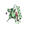
| ||||||||||||
| 3 | 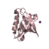
| ||||||||||||
| 4 | 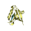
| ||||||||||||
| 5 | 
| ||||||||||||
| 6 | 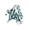
| ||||||||||||
| Unit cell |
|
-Components
| #1: Protein | Mass: 12709.242 Da / Num. of mol.: 6 Source method: isolated from a genetically manipulated source Source: (gene. exp.) Homo sapiens (human) / Gene: PTPN3, PTPH1 / Production host:  Escherichia coli BL21(DE3) (bacteria) / Strain (production host): B / References: UniProt: P26045, protein-tyrosine-phosphatase Escherichia coli BL21(DE3) (bacteria) / Strain (production host): B / References: UniProt: P26045, protein-tyrosine-phosphatase#2: Protein/peptide | Mass: 1393.554 Da / Num. of mol.: 6 Source method: isolated from a genetically manipulated source Source: (gene. exp.) Human papillomavirus type 16 / Gene: E6 / Production host: Escherichia coli BL21(DE3) (bacteria) / Strain (production host): B / References: UniProt: P03126#3: Chemical | ChemComp-IOD / Iodide  Mass: 126.904 Da / Num. of mol.: 6 / Source method: obtained synthetically / Formula: I Mass: 126.904 Da / Num. of mol.: 6 / Source method: obtained synthetically / Formula: I#4: Water | ChemComp-HOH / | Water Mass: 18.015 Da / Num. of mol.: 233 / Source method: isolated from a natural source / Formula: H2O Mass: 18.015 Da / Num. of mol.: 233 / Source method: isolated from a natural source / Formula: H2O |
|---|
-Experimental details
-Experiment
| Experiment | Method: X-RAY DIFFRACTION / Number of used crystals: 1 |
|---|
- Sample preparation
| Crystal | Density Matthews: 3.47 Å3/Da / Density % sol: 64.5 % |
|---|---|
| Crystal grow | Temperature: 277 K / Method: vapor diffusion, sitting drop / Details: 20% w/v PEG 3350 0.2 mM KI pH 7 |
-Data collection
| Diffraction | Mean temperature: 100 K |
|---|---|
| Diffraction source | Source: SYNCHROTRON / Site: SOLEIL  / Beamline: PROXIMA 2 / Wavelength: 0.978549599648 Å / Beamline: PROXIMA 2 / Wavelength: 0.978549599648 Å |
| Detector | Type: DECTRIS EIGER X 9M / Detector: PIXEL / Date: Feb 21, 2018 |
| Radiation | Protocol: SINGLE WAVELENGTH / Monochromatic (M) / Laue (L): M / Scattering type: x-ray |
| Radiation wavelength | Wavelength: 0.978549599648 Å / Relative weight: 1 |
| Reflection | Resolution: 2.19→46.62 Å / Num. obs: 47024 / % possible obs: 99.1 % / Redundancy: 6.9 % / Biso Wilson estimate: 47.643622563 Å2 / CC1/2: 0.996 / Rrim(I) all: 0.14 / Net I/σ(I): 8.17 |
| Reflection shell | Resolution: 2.19→2.23 Å / Redundancy: 6.5 % / Mean I/σ(I) obs: 1.11 / CC1/2: 0.726 / % possible all: 95.7 |
- Processing
| Software |
| ||||||||||||||||||||||||||||||||||||||||||||||||||||||||||||||||||||||||||||||||||||||||||||||||||||||||||||||||||||||||||||||
|---|---|---|---|---|---|---|---|---|---|---|---|---|---|---|---|---|---|---|---|---|---|---|---|---|---|---|---|---|---|---|---|---|---|---|---|---|---|---|---|---|---|---|---|---|---|---|---|---|---|---|---|---|---|---|---|---|---|---|---|---|---|---|---|---|---|---|---|---|---|---|---|---|---|---|---|---|---|---|---|---|---|---|---|---|---|---|---|---|---|---|---|---|---|---|---|---|---|---|---|---|---|---|---|---|---|---|---|---|---|---|---|---|---|---|---|---|---|---|---|---|---|---|---|---|---|---|---|
| Refinement | Method to determine structure: MOLECULAR REPLACEMENT Starting model: 5EZ0 Resolution: 2.19441072073→43.3432039427 Å / SU ML: 0.386490308239 / Cross valid method: FREE R-VALUE / σ(F): 1.36457088824 / Phase error: 32.2236856577
| ||||||||||||||||||||||||||||||||||||||||||||||||||||||||||||||||||||||||||||||||||||||||||||||||||||||||||||||||||||||||||||||
| Solvent computation | Shrinkage radii: 0.9 Å / VDW probe radii: 1.11 Å | ||||||||||||||||||||||||||||||||||||||||||||||||||||||||||||||||||||||||||||||||||||||||||||||||||||||||||||||||||||||||||||||
| Displacement parameters | Biso mean: 56.9100392383 Å2 | ||||||||||||||||||||||||||||||||||||||||||||||||||||||||||||||||||||||||||||||||||||||||||||||||||||||||||||||||||||||||||||||
| Refinement step | Cycle: LAST / Resolution: 2.19441072073→43.3432039427 Å
| ||||||||||||||||||||||||||||||||||||||||||||||||||||||||||||||||||||||||||||||||||||||||||||||||||||||||||||||||||||||||||||||
| Refine LS restraints |
| ||||||||||||||||||||||||||||||||||||||||||||||||||||||||||||||||||||||||||||||||||||||||||||||||||||||||||||||||||||||||||||||
| LS refinement shell |
| ||||||||||||||||||||||||||||||||||||||||||||||||||||||||||||||||||||||||||||||||||||||||||||||||||||||||||||||||||||||||||||||
| Refinement TLS params. | Method: refined / Origin x: -0.439637573517 Å / Origin y: 41.323561443 Å / Origin z: 32.3646606666 Å
| ||||||||||||||||||||||||||||||||||||||||||||||||||||||||||||||||||||||||||||||||||||||||||||||||||||||||||||||||||||||||||||||
| Refinement TLS group | Selection details: all |
