 Movie
Movie Controller

[English] 日本語
 Yorodumi
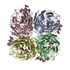
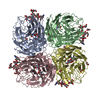
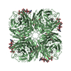
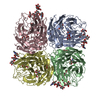
- PDB-6hgb: Influenza A virus N6 neuraminidase native structure (Duck/England/56). -
+ Open data

- Basic information
| Entry | Database: PDB / ID: 6hgb | |||||||||
|---|---|---|---|---|---|---|---|---|---|---|
| Title | Influenza A virus N6 neuraminidase native structure (Duck/England/56). | |||||||||
 Components Components | Neuraminidase | |||||||||
 Keywords Keywords | HYDROLASE / Influenza / A / Native / N6 / Neuraminidase / Duck / Virus / Enzyme / Complex / England / Wild type | |||||||||
| Function / homology |  Function and homology information Function and homology informationexo-alpha-(2->3)-sialidase activity / exo-alpha-(2->6)-sialidase activity / exo-alpha-(2->8)-sialidase activity / exo-alpha-sialidase / viral budding from plasma membrane / carbohydrate metabolic process / host cell plasma membrane / virion membrane / membrane / metal ion bindingSimilarity search - Function | |||||||||
| Biological species |   Influenza A virus Influenza A virus | |||||||||
| Method | X-RAY DIFFRACTION / SYNCHROTRON / MOLECULAR REPLACEMENT / Resolution: 1.5 Å | |||||||||
 Authors Authors | Salinger, M.T. / Hobbs, J.R. / Murray, J.W. / Laver, W.G. / Kuhn, P. / Garman, E.F. | |||||||||
| Funding support |  United Kingdom, 1items United Kingdom, 1items
| |||||||||
 Citation Citation | Journal: To Be Published Title: High Resolution Structures of Viral Neuraminidase with Drugs Bound in the Active Site. (In preparation) Authors: Salinger, M.T. / Hobbs, J.R. / Murray, J.W. / Laver, W.G. / Kuhn, P. / Garman, E.F. | |||||||||
| History |
|
- Structure visualization
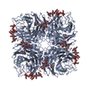
| Structure viewer | Molecule: MolmilJmol/JSmol |
|---|
- Downloads & links
-Download
| PDBx/mmCIF format | 6hgb.cif.gz | 382.3 KB | Display | PDBx/mmCIF format |
|---|---|---|---|---|
| PDB format | pdb6hgb.ent.gz | 312.1 KB | Display | PDB format |
| PDBx/mmJSON format | 6hgb.json.gz | Tree view | PDBx/mmJSON format | |
| Others |  Other downloads Other downloads |
-Validation report
| Arichive directory | https://data.pdbj.org/pub/pdb/validation_reports/hg/6hgbftp://data.pdbj.org/pub/pdb/validation_reports/hg/6hgb | HTTPS FTP |
|---|
-Related structure data
| Related structure data | 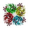 6hcxC  6hfcC 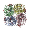 6hfyC 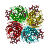 6hg0C 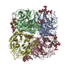 1v0zS S: Starting model for refinement C: citing same article ( |
|---|---|
| Similar structure data |


-Links
 PDBj
PDBj

- Assembly
| Deposited unit | 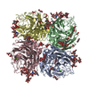
| ||||||||
|---|---|---|---|---|---|---|---|---|---|
| 1 |
| ||||||||
| Unit cell |
|
-Components
-Protein , 1 types, 4 molecules ABCD
| #1: Protein | Mass: 43011.531 Da / Num. of mol.: 4 / Source method: isolated from a natural source Source: (natural) Influenza A virus (strain A/Duck/England/1/1956 H11N6)Strain: A/Duck/England/1/1956 H11N6 / References: UniProt: Q6XV27, exo-alpha-sialidase |
|---|
-Sugars , 5 types, 12 molecules 
| #2: Polysaccharide | 2-acetamido-2-deoxy-beta-D-glucopyranose-(1-4)-2-acetamido-2-deoxy-beta-D-glucopyranose / Mass: 424.401 Da / Num. of mol.: 6Source method: isolated from a genetically manipulated source #3: Polysaccharide | alpha-D-mannopyranose-(1-2)-alpha-D-mannopyranose-(1-3)-[alpha-D-mannopyranose-(1-3)-[alpha-D- ...alpha-D-mannopyranose-(1-2)-alpha-D-mannopyranose-(1-3)-[alpha-D-mannopyranose-(1-3)-[alpha-D-mannopyranose-(1-6)]alpha-D-mannopyranose-(1-6)]beta-D-mannopyranose-(1-4)-2-acetamido-2-deoxy-beta-D-glucopyranose-(1-4)-2-acetamido-2-deoxy-beta-D-glucopyranose | / Mass: 1397.245 Da / Num. of mol.: 1Source method: isolated from a genetically manipulated source #4: Polysaccharide | alpha-D-mannopyranose-(1-3)-[alpha-D-mannopyranose-(1-6)]alpha-D-mannopyranose-(1-6)-[alpha-D- ...alpha-D-mannopyranose-(1-3)-[alpha-D-mannopyranose-(1-6)]alpha-D-mannopyranose-(1-6)-[alpha-D-mannopyranose-(1-3)]beta-D-mannopyranose-(1-4)-2-acetamido-2-deoxy-beta-D-glucopyranose-(1-4)-2-acetamido-2-deoxy-beta-D-glucopyranose | / Mass: 1235.105 Da / Num. of mol.: 1Source method: isolated from a genetically manipulated source #5: Polysaccharide | / Mass: 1072.964 Da / Num. of mol.: 2Source method: isolated from a genetically manipulated source #10: Sugar | N-Acetylglucosamine Type: D-saccharide, beta linking / Mass: 221.208 Da / Num. of mol.: 2 Type: D-saccharide, beta linking / Mass: 221.208 Da / Num. of mol.: 2Source method: isolated from a genetically manipulated source Formula: C8H15NO6 |
|---|
-Non-polymers , 5 types, 1638 molecules 


| #6: Chemical | ChemComp-GOL / Glycerol Mass: 92.094 Da / Num. of mol.: 25 / Source method: isolated from a natural source / Formula: C3H8O3 Mass: 92.094 Da / Num. of mol.: 25 / Source method: isolated from a natural source / Formula: C3H8O3#7: Chemical | ChemComp-CA /  Mass: 40.078 Da / Num. of mol.: 4 / Source method: obtained synthetically / Formula: Ca Mass: 40.078 Da / Num. of mol.: 4 / Source method: obtained synthetically / Formula: Ca#8: Chemical | Phosphate Mass: 94.971 Da / Num. of mol.: 3 / Source method: isolated from a natural source / Formula: PO4 Mass: 94.971 Da / Num. of mol.: 3 / Source method: isolated from a natural source / Formula: PO4#9: Chemical | ChemComp-PEG / Diethylene glycol Mass: 106.120 Da / Num. of mol.: 19 / Source method: obtained synthetically / Formula: C4H10O3 Mass: 106.120 Da / Num. of mol.: 19 / Source method: obtained synthetically / Formula: C4H10O3#11: Water | ChemComp-HOH / | WaterMass: 18.015 Da / Num. of mol.: 1587 / Source method: isolated from a natural source / Formula: H2O |
|---|
-Experimental details
-Experiment
| Experiment | Method: X-RAY DIFFRACTION / Number of used crystals: 1 |
|---|
- Sample preparation
| Crystal | Density Matthews: 2.43 Å3/Da / Density % sol: 49.32 % |
|---|---|
| Crystal grow | Temperature: 293 K / Method: vapor diffusion, hanging drop Details: N6 crystals were grown by hanging-drop vapour diffusion against 1:1 20%(w/v) PEG-3350 and 2 microM CaCl2 in 150mM NaCl at 20 degrees celsius, starting with equal volumes of N6 (20mg/ml in ...Details: N6 crystals were grown by hanging-drop vapour diffusion against 1:1 20%(w/v) PEG-3350 and 2 microM CaCl2 in 150mM NaCl at 20 degrees celsius, starting with equal volumes of N6 (20mg/ml in saline) and 20% (w/v) PEG 3350 and 2 microM in 1% saline. Microseeding with crystals of native N6 was carried out using a super saturated solution of N6 NA in 20% PEG 3350. Temp details: Kept constant throughout. |
-Data collection
| Diffraction | Mean temperature: 100 K |
|---|---|
| Diffraction source | Source: SYNCHROTRON / Site: SSRL  / Beamline: BL9-1 / Wavelength: 0.93 Å / Beamline: BL9-1 / Wavelength: 0.93 Å |
| Detector | Type: MAR scanner 345 mm plate / Detector: IMAGE PLATE / Date: Dec 6, 1999 |
| Radiation | Protocol: SINGLE WAVELENGTH / Monochromatic (M) / Laue (L): M / Scattering type: x-ray |
| Radiation wavelength | Wavelength: 0.93 Å / Relative weight: 1 |
| Reflection | Resolution: 1.5→30.68 Å / Num. obs: 241948 / % possible obs: 92.1 % / Redundancy: 3.7 % / Rsym value: 0.085 / Net I/σ(I): 9.5 |
| Reflection shell | Resolution: 1.5→1.52 Å / Mean I/σ(I) obs: 2.7 / Num. unique obs: 10672 / Rsym value: 0.184 / % possible all: 82.5 |
- Processing
| Software |
| ||||||||||||||||||||||||||||||||||||||||||||||||||||||||||||||||||||||||||||||||||||||||||||||||||||||||||||||||||||||||||||||||||||||||||||||||||||||||||||||||||||||||||||||||||||||
|---|---|---|---|---|---|---|---|---|---|---|---|---|---|---|---|---|---|---|---|---|---|---|---|---|---|---|---|---|---|---|---|---|---|---|---|---|---|---|---|---|---|---|---|---|---|---|---|---|---|---|---|---|---|---|---|---|---|---|---|---|---|---|---|---|---|---|---|---|---|---|---|---|---|---|---|---|---|---|---|---|---|---|---|---|---|---|---|---|---|---|---|---|---|---|---|---|---|---|---|---|---|---|---|---|---|---|---|---|---|---|---|---|---|---|---|---|---|---|---|---|---|---|---|---|---|---|---|---|---|---|---|---|---|---|---|---|---|---|---|---|---|---|---|---|---|---|---|---|---|---|---|---|---|---|---|---|---|---|---|---|---|---|---|---|---|---|---|---|---|---|---|---|---|---|---|---|---|---|---|---|---|---|---|
| Refinement | Method to determine structure: MOLECULAR REPLACEMENT Starting model: 1V0Z Resolution: 1.5→30.68 Å / Cor.coef. Fo:Fc: 0.976 / Cor.coef. Fo:Fc free: 0.966 / SU B: 1.556 / SU ML: 0.054 / Cross valid method: THROUGHOUT / ESU R: 0.065 / ESU R Free: 0.065 / Stereochemistry target values: MAXIMUM LIKELIHOOD / Details: HYDROGENS HAVE BEEN ADDED IN THE RIDING POSITIONS
| ||||||||||||||||||||||||||||||||||||||||||||||||||||||||||||||||||||||||||||||||||||||||||||||||||||||||||||||||||||||||||||||||||||||||||||||||||||||||||||||||||||||||||||||||||||||
| Solvent computation | Ion probe radii: 0.8 Å / Shrinkage radii: 0.8 Å / VDW probe radii: 1.2 Å / Solvent model: MASK | ||||||||||||||||||||||||||||||||||||||||||||||||||||||||||||||||||||||||||||||||||||||||||||||||||||||||||||||||||||||||||||||||||||||||||||||||||||||||||||||||||||||||||||||||||||||
| Displacement parameters | Biso mean: 13.965 Å2
| ||||||||||||||||||||||||||||||||||||||||||||||||||||||||||||||||||||||||||||||||||||||||||||||||||||||||||||||||||||||||||||||||||||||||||||||||||||||||||||||||||||||||||||||||||||||
| Refinement step | Cycle: 1 / Resolution: 1.5→30.68 Å
| ||||||||||||||||||||||||||||||||||||||||||||||||||||||||||||||||||||||||||||||||||||||||||||||||||||||||||||||||||||||||||||||||||||||||||||||||||||||||||||||||||||||||||||||||||||||
| Refine LS restraints |
|