 Movie
Movie Controller

[English] 日本語
 Yorodumi
- PDB-6h4g: Regulatory subunit of a cAMP-independent protein kinase A from Tr... -
+ Open data

- Basic information
| Entry | Database: PDB / ID: 6h4g | |||||||||
|---|---|---|---|---|---|---|---|---|---|---|
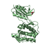
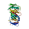
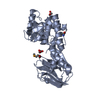
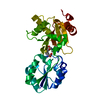
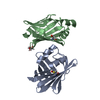
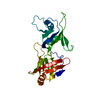
| Title | Regulatory subunit of a cAMP-independent protein kinase A from Trypanosoma brucei: E311A, T318R, V319A mutant bound to cAMP in the A site | |||||||||
 Components Components | Protein kinase A regulatory subunit | |||||||||
 Keywords Keywords |  SIGNALING PROTEIN / Protein kinase A / Regulatory Subunit / Nucleoside-binding protein SIGNALING PROTEIN / Protein kinase A / Regulatory Subunit / Nucleoside-binding protein | |||||||||
| Function / homology |  Function and homology information Function and homology informationmodulation of formation of structure involved in a symbiotic process / cilium movement involved in cell motility / cAMP-dependent protein kinase regulator activity / cAMP-dependent protein kinase complex / cilium / kinase activity / intracellular signal transduction / protein phosphorylationSimilarity search - Function | |||||||||
| Biological species |  Trypanosoma brucei brucei TREU927 (eukaryote) Trypanosoma brucei brucei TREU927 (eukaryote) | |||||||||
| Method | X-RAY DIFFRACTION / SYNCHROTRON / MOLECULAR REPLACEMENT / Resolution: 2.14351698698 Å | |||||||||
 Authors Authors | Volpato Santos, Y. / Lorentzen, E. / Basquin, J. / Boshart, M. | |||||||||
| Funding support |  Brazil, Brazil,  Germany, 2items Germany, 2items
| |||||||||
 Citation Citation | Journal: Elife / Year: 2024 Title: Purine nucleosides replace cAMP in allosteric regulation of PKA in trypanosomatid pathogens. Authors: Ober, V.T. / Githure, G.B. / Volpato Santos, Y. / Becker, S. / Moya Munoz, G. / Basquin, J. / Schwede, F. / Lorentzen, E. / Boshart, M. | |||||||||
| History |
|
- Structure visualization
| Structure viewer | Molecule: MolmilJmol/JSmol |
|---|
- Downloads & links
-Download
| PDBx/mmCIF format | 6h4g.cif.gz | 289.6 KB | Display | PDBx/mmCIF format |
|---|---|---|---|---|
| PDB format | pdb6h4g.ent.gz | 193.1 KB | Display | PDB format |
| PDBx/mmJSON format | 6h4g.json.gz | Tree view | PDBx/mmJSON format | |
| Others |  Other downloads Other downloads |
-Validation report
| Arichive directory | https://data.pdbj.org/pub/pdb/validation_reports/h4/6h4gftp://data.pdbj.org/pub/pdb/validation_reports/h4/6h4g | HTTPS FTP |
|---|
-Related structure data
| Related structure data |  6floSC  6hyiC S: Starting model for refinement C: citing same article ( |
|---|---|
| Similar structure data |


-Links
 PDBj
PDBj


- Assembly
| Deposited unit | 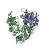
| ||||||||||||
|---|---|---|---|---|---|---|---|---|---|---|---|---|---|
| 1 | 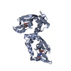
| ||||||||||||
| 2 | 
| ||||||||||||
| Unit cell |
|
-Components
| #1: Protein | Mass: 34383.027 Da / Num. of mol.: 2 / Mutation: E311A, T318R, V319A Source method: isolated from a genetically manipulated source Source: (gene. exp.) Trypanosoma brucei brucei TREU927 (eukaryote)Gene: Tb11.02.2210 Production host:  Escherichia coli 'BL21-Gold(DE3)pLysS AG' (bacteria) Escherichia coli 'BL21-Gold(DE3)pLysS AG' (bacteria)References: UniProt: Q385V6 #2: Chemical | Cyclic adenosine monophosphate  Mass: 329.206 Da / Num. of mol.: 2 / Source method: obtained synthetically / Formula: C10H12N5O6P Mass: 329.206 Da / Num. of mol.: 2 / Source method: obtained synthetically / Formula: C10H12N5O6P#3: Chemical | Inosine  Mass: 268.226 Da / Num. of mol.: 2 / Source method: obtained synthetically / Formula: C10H12N4O5 Mass: 268.226 Da / Num. of mol.: 2 / Source method: obtained synthetically / Formula: C10H12N4O5#4: Water | ChemComp-HOH / | Water Mass: 18.015 Da / Num. of mol.: 320 / Source method: isolated from a natural source / Formula: H2O Mass: 18.015 Da / Num. of mol.: 320 / Source method: isolated from a natural source / Formula: H2O |
|---|
-Experimental details
-Experiment
| Experiment | Method: X-RAY DIFFRACTION / Number of used crystals: 1 |
|---|
- Sample preparation
| Crystal | Density Matthews: 2.24 Å3/Da / Density % sol: 45.09 % |
|---|---|
| Crystal grow | Temperature: 277 K / Method: vapor diffusion, sitting drop Details: 50 mM Tris pH 8.0 0.2 mM Magnesium Chloride 40% PEG 3350 |
-Data collection
| Diffraction | Mean temperature: 100 K |
|---|---|
| Diffraction source | Source: SYNCHROTRON / Site: SLS  / Beamline: X06DA / Wavelength: 1 Å / Beamline: X06DA / Wavelength: 1 Å |
| Detector | Type: DECTRIS PILATUS 2M / Detector: PIXEL / Date: Nov 19, 2016 |
| Radiation | Protocol: SINGLE WAVELENGTH / Monochromatic (M) / Laue (L): M / Scattering type: x-ray |
| Radiation wavelength | Wavelength: 1 Å / Relative weight: 1 |
| Reflection | Resolution: 2.144→30.21 Å / Num. obs: 31343 / % possible obs: 100 % / Redundancy: 1 % / Biso Wilson estimate: 17.8216436493 Å2 / Rpim(I) all: 0.0888 / Net I/σ(I): 0.92 |
| Reflection shell | Resolution: 2.144→2.24 Å / Rpim(I) all: 0.1223 |
- Processing
| Software |
| |||||||||||||||||||||||||||||||||||||||||||||||||||||||||||||||||||||||||||||||||||||||||||||||||||||||||
|---|---|---|---|---|---|---|---|---|---|---|---|---|---|---|---|---|---|---|---|---|---|---|---|---|---|---|---|---|---|---|---|---|---|---|---|---|---|---|---|---|---|---|---|---|---|---|---|---|---|---|---|---|---|---|---|---|---|---|---|---|---|---|---|---|---|---|---|---|---|---|---|---|---|---|---|---|---|---|---|---|---|---|---|---|---|---|---|---|---|---|---|---|---|---|---|---|---|---|---|---|---|---|---|---|---|---|
| Refinement | Method to determine structure: MOLECULAR REPLACEMENT Starting model: 6FLO Resolution: 2.14351698698→30.205523826 Å / SU ML: 0.380726826369 / Cross valid method: FREE R-VALUE / σ(F): 1.34684365027 / Phase error: 31.9468288459
| |||||||||||||||||||||||||||||||||||||||||||||||||||||||||||||||||||||||||||||||||||||||||||||||||||||||||
| Solvent computation | Shrinkage radii: 0.9 Å / VDW probe radii: 1.11 Å | |||||||||||||||||||||||||||||||||||||||||||||||||||||||||||||||||||||||||||||||||||||||||||||||||||||||||
| Displacement parameters | Biso mean: 21.8229744064 Å2 | |||||||||||||||||||||||||||||||||||||||||||||||||||||||||||||||||||||||||||||||||||||||||||||||||||||||||
| Refinement step | Cycle: LAST / Resolution: 2.14351698698→30.205523826 Å
| |||||||||||||||||||||||||||||||||||||||||||||||||||||||||||||||||||||||||||||||||||||||||||||||||||||||||
| Refine LS restraints |
| |||||||||||||||||||||||||||||||||||||||||||||||||||||||||||||||||||||||||||||||||||||||||||||||||||||||||
| LS refinement shell |
| |||||||||||||||||||||||||||||||||||||||||||||||||||||||||||||||||||||||||||||||||||||||||||||||||||||||||
| Refinement TLS params. | Method: refined / Origin x: -20.3825618882 Å / Origin y: -12.350172076 Å / Origin z: 23.2982273412 Å
| |||||||||||||||||||||||||||||||||||||||||||||||||||||||||||||||||||||||||||||||||||||||||||||||||||||||||
| Refinement TLS group | Selection details: all |
