 Movie
Movie Controller

[English] 日本語
 Yorodumi
- PDB-6h1y: CRYSTAL STRUCTURE OF A CHIMERIC VARIANT OF THIOREDOXIN FROM ESCHE... -
+ Open data

- Basic information
| Entry | Database: PDB / ID: 6h1y | ||||||
|---|---|---|---|---|---|---|---|
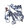
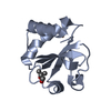
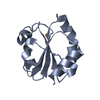
| Title | CRYSTAL STRUCTURE OF A CHIMERIC VARIANT OF THIOREDOXIN FROM ESCHERICHIA COLI | ||||||
 Components Components | Thioredoxin 1,Thioredoxin (TrxA-1),Thioredoxin 1 | ||||||
 Keywords Keywords |  OXIDOREDUCTASE / protein thermal stability / loop size optimization / fundamental rules / chimeric proteins / protein stabilization OXIDOREDUCTASE / protein thermal stability / loop size optimization / fundamental rules / chimeric proteins / protein stabilization | ||||||
| Function / homology |  Function and homology information Function and homology informationDNA polymerase processivity factor activity / protein-disulfide reductase activity / cell redox homeostasis / cytosol / cytoplasmSimilarity search - Function | ||||||
| Biological species |  Escherichia coli (E. coli) Escherichia coli (E. coli)  Sulfolobus solfataricus (archaea) Sulfolobus solfataricus (archaea) | ||||||
| Method | X-RAY DIFFRACTION / MOLECULAR REPLACEMENT / Resolution: 2.99 Å | ||||||
 Authors Authors | Ruggiero, A. / Smaldone, G. / Esposito, L. / Balasco, N. / Vitagliano, L. | ||||||
 Citation Citation | Journal: Febs J. / Year: 2019 Title: Loop size optimization induces a strong thermal stabilization of the thioredoxin fold. Authors: Ruggiero, A. / Smaldone, G. / Esposito, L. / Balasco, N. / Vitagliano, L. | ||||||
| History |
|
- Structure visualization
| Structure viewer | Molecule: MolmilJmol/JSmol |
|---|
- Downloads & links
-Download
| PDBx/mmCIF format | 6h1y.cif.gz | 53.4 KB | Display | PDBx/mmCIF format |
|---|---|---|---|---|
| PDB format | pdb6h1y.ent.gz | 38.3 KB | Display | PDB format |
| PDBx/mmJSON format | 6h1y.json.gz | Tree view | PDBx/mmJSON format | |
| Others |  Other downloads Other downloads |
-Validation report
| Arichive directory | https://data.pdbj.org/pub/pdb/validation_reports/h1/6h1yftp://data.pdbj.org/pub/pdb/validation_reports/h1/6h1y | HTTPS FTP |
|---|
-Related structure data
| Related structure data | 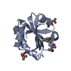 2trxS S: Starting model for refinement |
|---|---|
| Similar structure data |


-Links
 PDBj
PDBj

- Assembly
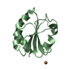
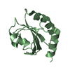
| Deposited unit | 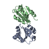
| ||||||||
|---|---|---|---|---|---|---|---|---|---|
| 1 | 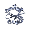
| ||||||||
| 2 | 
| ||||||||
| Unit cell |
|
-Components
| #1: Protein | Mass: 11784.570 Da / Num. of mol.: 2 Source method: isolated from a genetically manipulated source Source: (gene. exp.) Escherichia coli (E. coli), (gene. exp.) Sulfolobus solfataricus (archaea)Strain: K12, ATCC 35092 / DSM 1617 / JCM 11322 / P2 / Gene: trxA, fipA, tsnC, b3781, JW5856, trxA-1, SSO0368 / Production host: Escherichia coli (E. coli) / References: UniProt: P0AA25, UniProt: Q980E5 |
|---|
-Experimental details
-Experiment
| Experiment | Method: X-RAY DIFFRACTION / Number of used crystals: 1 |
|---|
- Sample preparation
| Crystal | Density Matthews: 4.41 Å3/Da / Density % sol: 72.13 % |
|---|---|
| Crystal grow | Temperature: 293 K / Method: vapor diffusion, hanging drop Details: Crystals were obtained with a protein concentration of 20-25 mg/ml. The composition of the reservoir solution was 17% (w/v) PEG10000, 0.1 M ammonium acetate and 0.1 M BIS-TRIS buffer, pH 5.5. |
-Data collection
| Diffraction | Mean temperature: 100 K |
|---|---|
| Diffraction source | Source: ROTATING ANODE / Type: RIGAKU MICROMAX-007 / Wavelength: 1.54 Å |
| Detector | Type: RIGAKU SATURN 944 / Detector: CCD / Date: Feb 19, 2014 |
| Radiation | Protocol: SINGLE WAVELENGTH / Monochromatic (M) / Laue (L): M / Scattering type: x-ray |
| Radiation wavelength | Wavelength: 1.54 Å / Relative weight: 1 |
| Reflection | Resolution: 2.99→82.57 Å / Num. obs: 7669 / % possible obs: 93.6 % / Redundancy: 2.7 % / Rmerge(I) obs: 0.079 / Net I/σ(I): 14.8 |
| Reflection shell | Resolution: 2.99→3.13 Å |
- Processing
| Software |
| ||||||||||||||||||||||||||||||||||||||||||||||||||||||||||||||||||||||||||||||||||||||||||||||||||||||||||||||||||||||||||||||||||||||||||||||||||||||||||||||||||||||||||||||||||||||
|---|---|---|---|---|---|---|---|---|---|---|---|---|---|---|---|---|---|---|---|---|---|---|---|---|---|---|---|---|---|---|---|---|---|---|---|---|---|---|---|---|---|---|---|---|---|---|---|---|---|---|---|---|---|---|---|---|---|---|---|---|---|---|---|---|---|---|---|---|---|---|---|---|---|---|---|---|---|---|---|---|---|---|---|---|---|---|---|---|---|---|---|---|---|---|---|---|---|---|---|---|---|---|---|---|---|---|---|---|---|---|---|---|---|---|---|---|---|---|---|---|---|---|---|---|---|---|---|---|---|---|---|---|---|---|---|---|---|---|---|---|---|---|---|---|---|---|---|---|---|---|---|---|---|---|---|---|---|---|---|---|---|---|---|---|---|---|---|---|---|---|---|---|---|---|---|---|---|---|---|---|---|---|---|
| Refinement | Method to determine structure: MOLECULAR REPLACEMENT Starting model: 2TRX Resolution: 2.99→82.57 Å / Cor.coef. Fo:Fc: 0.933 / Cor.coef. Fo:Fc free: 0.893 / SU B: 18.35 / SU ML: 0.323 / Cross valid method: THROUGHOUT / ESU R: 3.049 / ESU R Free: 0.412 / Details: HYDROGENS HAVE BEEN ADDED IN THE RIDING POSITIONS
| ||||||||||||||||||||||||||||||||||||||||||||||||||||||||||||||||||||||||||||||||||||||||||||||||||||||||||||||||||||||||||||||||||||||||||||||||||||||||||||||||||||||||||||||||||||||
| Solvent computation | Ion probe radii: 0.8 Å / Shrinkage radii: 0.8 Å / VDW probe radii: 1.2 Å | ||||||||||||||||||||||||||||||||||||||||||||||||||||||||||||||||||||||||||||||||||||||||||||||||||||||||||||||||||||||||||||||||||||||||||||||||||||||||||||||||||||||||||||||||||||||
| Displacement parameters | Biso mean: 53.326 Å2
| ||||||||||||||||||||||||||||||||||||||||||||||||||||||||||||||||||||||||||||||||||||||||||||||||||||||||||||||||||||||||||||||||||||||||||||||||||||||||||||||||||||||||||||||||||||||
| Refinement step | Cycle: 1 / Resolution: 2.99→82.57 Å
| ||||||||||||||||||||||||||||||||||||||||||||||||||||||||||||||||||||||||||||||||||||||||||||||||||||||||||||||||||||||||||||||||||||||||||||||||||||||||||||||||||||||||||||||||||||||
| Refine LS restraints |
|