 Movie
Movie Controller


+ Open data

- Basic information
| Entry | Database: PDB / ID: 6gzw | ||||||
|---|---|---|---|---|---|---|---|
| Title | Ferric DtpA from Streptomyces lividans | ||||||
 Components Components | Dye type peroxidase A | ||||||
 Keywords Keywords |  OXIDOREDUCTASE / peroxidase / ferric / dye-type OXIDOREDUCTASE / peroxidase / ferric / dye-type | ||||||
| Function / homology |  Function and homology information Function and homology informationiron import into cell / Oxidoreductases; Acting on a peroxide as acceptor; Peroxidases / peroxidase activity / heme binding / metal ion bindingSimilarity search - Function | ||||||
| Biological species |  Streptomyces lividans 1326 (bacteria) Streptomyces lividans 1326 (bacteria) | ||||||
| Method | X-RAY DIFFRACTION / SYNCHROTRON / MOLECULAR REPLACEMENT / Resolution: 1.41 Å | ||||||
 Authors Authors | Moreno Chicano, T. / Worrall, J.A.R. / Hough, M.A. | ||||||
| Funding support |  United Kingdom, 1items United Kingdom, 1items
| ||||||
 Citation Citation | Journal: Chemistry / Year: 2019 Title: An Aromatic Dyad Motif in Dye Decolourising Peroxidases Has Implications for Free Radical Formation and Catalysis. Authors: Chaplin, A.K. / Chicano, T.M. / Hampshire, B.V. / Wilson, M.T. / Hough, M.A. / Svistunenko, D.A. / Worrall, J.A.R. | ||||||
| History |
|
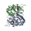
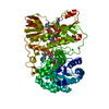
- Structure visualization
| Structure viewer | Molecule: MolmilJmol/JSmol |
|---|
- Downloads & links
-Download
| PDBx/mmCIF format | 6gzw.cif.gz | 174.6 KB | Display | PDBx/mmCIF format |
|---|---|---|---|---|
| PDB format | pdb6gzw.ent.gz | 134.3 KB | Display | PDB format |
| PDBx/mmJSON format | 6gzw.json.gz | Tree view | PDBx/mmJSON format | |
| Others |  Other downloads Other downloads |
-Validation report
| Arichive directory | https://data.pdbj.org/pub/pdb/validation_reports/gz/6gzwftp://data.pdbj.org/pub/pdb/validation_reports/gz/6gzw | HTTPS FTP |
|---|
-Related structure data
| Related structure data |  5mapS S: Starting model for refinement |
|---|---|
| Similar structure data |


-Links
 PDBj
PDBj

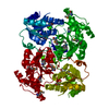
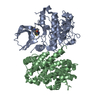
- Assembly
| Deposited unit | 
| ||||||||
|---|---|---|---|---|---|---|---|---|---|
| 1 |
| ||||||||
| Unit cell |
|
-Components
| #1: Protein | Mass: 40581.320 Da / Num. of mol.: 2 Source method: isolated from a genetically manipulated source Source: (gene. exp.) Streptomyces lividans 1326 (bacteria) / Plasmid: pet28a / Production host: Escherichia coli BL21 (bacteria) / References: UniProt: A0A076MAJ9#2: Chemical | Heme B  Mass: 616.487 Da / Num. of mol.: 2 / Source method: obtained synthetically / Formula: C34H32FeN4O4 Mass: 616.487 Da / Num. of mol.: 2 / Source method: obtained synthetically / Formula: C34H32FeN4O4#3: Water | ChemComp-HOH / | Water Mass: 18.015 Da / Num. of mol.: 793 / Source method: isolated from a natural source / Formula: H2O Mass: 18.015 Da / Num. of mol.: 793 / Source method: isolated from a natural source / Formula: H2O |
|---|
-Experimental details
-Experiment
| Experiment | Method: X-RAY DIFFRACTION / Number of used crystals: 1 |
|---|
- Sample preparation
| Crystal | Density Matthews: 2.09 Å3/Da / Density % sol: 41.01 % |
|---|---|
| Crystal grow | Temperature: 291 K / Method: vapor diffusion, hanging drop / pH: 5.5 Details: Crystals were grown by the vapour diffusion hanging drop method using a ferric protein solution at 13 mg/ml equilibrated against a reservoir consisting of 17-24% PEG 3000 and 50-100 mM sodium citrate, pH 5.5. |
-Data collection
| Diffraction | Mean temperature: 100 K |
|---|---|
| Diffraction source | Source: SYNCHROTRON / Site: ESRF  / Beamline: MASSIF-3 / Wavelength: 0.9677 Å / Beamline: MASSIF-3 / Wavelength: 0.9677 Å |
| Detector | Type: DECTRIS EIGER X 16M / Detector: PIXEL / Date: Dec 15, 2016 |
| Radiation | Protocol: SINGLE WAVELENGTH / Monochromatic (M) / Laue (L): M / Scattering type: x-ray |
| Radiation wavelength | Wavelength: 0.9677 Å / Relative weight: 1 |
| Reflection | Resolution: 1.41→77.99 Å / Num. obs: 118339 / % possible obs: 93.9 % / Redundancy: 5.2 % / CC1/2: 0.997 / Rmerge(I) obs: 0.127 / Rpim(I) all: 0.06 / Rrim(I) all: 0.141 / Net I/σ(I): 7.8 |
| Reflection shell | Resolution: 1.41→1.46 Å / Redundancy: 5.1 % / Rmerge(I) obs: 1.253 / Num. unique obs: 9905 / CC1/2: 0.529 / Rpim(I) all: 0.593 / Rrim(I) all: 1.391 / % possible all: 80.4 |
- Processing
| Software |
| ||||||||||||||||||||||||||||||||||||||||||||||||||||||||||||
|---|---|---|---|---|---|---|---|---|---|---|---|---|---|---|---|---|---|---|---|---|---|---|---|---|---|---|---|---|---|---|---|---|---|---|---|---|---|---|---|---|---|---|---|---|---|---|---|---|---|---|---|---|---|---|---|---|---|---|---|---|---|
| Refinement | Method to determine structure: MOLECULAR REPLACEMENT Starting model: 5MAP Resolution: 1.41→77.99 Å / Cor.coef. Fo:Fc: 0.965 / Cor.coef. Fo:Fc free: 0.955 / SU B: 1.757 / SU ML: 0.064 / SU R Cruickshank DPI: 0.0768 / Cross valid method: THROUGHOUT / σ(F): 0 / ESU R: 0.077 / ESU R Free: 0.077 Details: HYDROGENS HAVE BEEN ADDED IN THE RIDING POSITIONS U VALUES : REFINED INDIVIDUALLY
| ||||||||||||||||||||||||||||||||||||||||||||||||||||||||||||
| Solvent computation | Ion probe radii: 0.8 Å / Shrinkage radii: 0.8 Å / VDW probe radii: 1.2 Å | ||||||||||||||||||||||||||||||||||||||||||||||||||||||||||||
| Displacement parameters | Biso max: 48.47 Å2 / Biso mean: 15.64 Å2 / Biso min: 3.49 Å2
| ||||||||||||||||||||||||||||||||||||||||||||||||||||||||||||
| Refinement step | Cycle: final / Resolution: 1.41→77.99 Å
| ||||||||||||||||||||||||||||||||||||||||||||||||||||||||||||
| Refine LS restraints |
| ||||||||||||||||||||||||||||||||||||||||||||||||||||||||||||
| LS refinement shell | Resolution: 1.41→1.447 Å / Rfactor Rfree error: 0 / Total num. of bins used: 20
|
