- PDB-6gy2: Crystal structure of human Plk1-PBD in complex with WSSSLATPPTLSS... -
+
Open data
ID or keywords:
Loading...
-
Basic information
Entry
Database: PDB / ID: 6gy2
Title
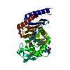
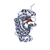
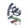
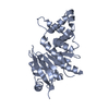
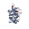
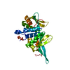
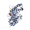
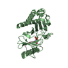
Crystal structure of human Plk1-PBD in complex with WSSSLATPPTLSSpTVLI phosphopeptide from BRCA2
Components
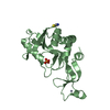
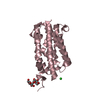
Phosphopeptide of BRCA2
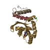
Serine/threonine-protein kinase PLK1
Keywords
CELL CYCLE / phosphorylation / chromosomes alignement / DNA repair
Function / homology
Function and homology information
BRCA2-MAGE-D1 complex / negative regulation of mammary gland epithelial cell proliferation / mitotic recombination-dependent replication fork processing / Mitotic Telophase/Cytokinesis / regulation of protein localization to cell cortex / Mitotic Metaphase/Anaphase Transition / Golgi inheritance / synaptonemal complex disassembly / establishment of protein localization to telomere / mitotic nuclear membrane disassembly ...BRCA2-MAGE-D1 complex / negative regulation of mammary gland epithelial cell proliferation / mitotic recombination-dependent replication fork processing / Mitotic Telophase/Cytokinesis / regulation of protein localization to cell cortex / Mitotic Metaphase/Anaphase Transition / Golgi inheritance / synaptonemal complex disassembly / establishment of protein localization to telomere / mitotic nuclear membrane disassembly / Activation of NIMA Kinases NEK9, NEK6, NEK7 / homologous chromosome segregation / protein localization to nuclear envelope / polo kinase / nuclear membrane disassembly / Impaired BRCA2 translocation to the nucleus / Impaired BRCA2 binding to SEM1 (DSS1) / Phosphorylation of Emi1 / nuclear ubiquitin ligase complex / metaphase/anaphase transition of mitotic cell cycle / histone H3 acetyltransferase activity / histone H4 acetyltransferase activity / synaptonemal complex / female meiosis chromosome segregation / lateral element / telomere maintenance via recombination / outer kinetochore / Phosphorylation of the APC/C / anaphase-promoting complex binding / regulation of protein binding / regulation of DNA damage checkpoint / double-strand break repair via alternative nonhomologous end joining / negative regulation of cyclin-dependent protein serine/threonine kinase activity / HDR through MMEJ (alt-NHEJ) / positive regulation of ubiquitin protein ligase activity / oocyte maturation / Impaired BRCA2 binding to PALB2 / regulation of mitotic spindle assembly / microtubule bundle formation / gamma-tubulin binding / Polo-like kinase mediated events / mitotic chromosome condensation / Golgi Cisternae Pericentriolar Stack Reorganization / DNA repair complex / positive regulation of ubiquitin-protein transferase activity / sister chromatid cohesion / regulation of mitotic metaphase/anaphase transition / centrosome cycle / Defective homologous recombination repair (HRR) due to BRCA1 loss of function / Defective HDR through Homologous Recombination Repair (HRR) due to PALB2 loss of BRCA1 binding function / Defective HDR through Homologous Recombination Repair (HRR) due to PALB2 loss of BRCA2/RAD51/RAD51C binding function / Homologous DNA Pairing and Strand Exchange / Resolution of D-loop Structures through Synthesis-Dependent Strand Annealing (SDSA) / response to UV-C / Resolution of D-loop Structures through Holliday Junction Intermediates / regulation of mitotic cell cycle phase transition / inner cell mass cell proliferation / Impaired BRCA2 binding to RAD51 / DNA damage response, signal transduction by p53 class mediator resulting in transcription of p21 class mediator / hematopoietic stem cell proliferation / female gonad development / mitotic spindle assembly checkpoint signaling / mitotic spindle pole / male meiosis I / regulation of mitotic cell cycle / mitotic sister chromatid segregation / regulation of anaphase-promoting complex-dependent catabolic process / mitotic G2 DNA damage checkpoint signaling / positive regulation of proteolysis / establishment of mitotic spindle orientation / Presynaptic phase of homologous DNA pairing and strand exchange / centrosome duplication / mitotic cytokinesis / centriolar satellite / response to X-ray / intrinsic apoptotic signaling pathway in response to DNA damage by p53 class mediator / spindle midzone / Amplification of signal from unattached kinetochores via a MAD2 inhibitory signal / Cyclin A/B1/B2 associated events during G2/M transition / Mitotic Prometaphase / EML4 and NUDC in mitotic spindle formation / Loss of Nlp from mitotic centrosomes / Loss of proteins required for interphase microtubule organization from the centrosome / Recruitment of mitotic centrosome proteins and complexes / protein localization to chromatin / Resolution of Sister Chromatid Cohesion / Recruitment of NuMA to mitotic centrosomes / Anchoring of the basal body to the plasma membrane / centriole / positive regulation of mitotic cell cycle / AURKA Activation by TPX2 / mitotic spindle organization / Condensation of Prophase Chromosomes / positive regulation of peptidyl-threonine phosphorylation / regulation of cytokinesis / secretory granule / RHO GTPases Activate Formins / cellular response to ionizing radiation / nucleotide-excision repair / response to gamma radiation Similarity search - Function
: / BRCA2, OB2 / BRCA2, OB3 / Tower domain / Breast cancer type 2 susceptibility protein, helical domain / BRCA2 helical domain superfamily / BRCA2, oligonucleotide/oligosaccharide-binding, domain 3 / Tower / BRCA2, helical / Tower ...: / BRCA2, OB2 / BRCA2, OB3 / Tower domain / Breast cancer type 2 susceptibility protein, helical domain / BRCA2 helical domain superfamily / BRCA2, oligonucleotide/oligosaccharide-binding, domain 3 / Tower / BRCA2, helical / Tower / BRCA2 repeat / BRCA2, OB1 / Breast cancer type 2 susceptibility protein / BRCA2 repeat / BRCA2, oligonucleotide/oligosaccharide-binding, domain 1 / BRCA2 repeat profile. / POLO box domain / Polo-like kinase 1, catalytic domain / Second polo-box domain / First polo-box domain / POLO box domain superfamily / POLO box duplicated region / POLO box domain / POLO box domain profile. / Arylsulfatase, C-terminal domain / Serine/threonine-protein kinase, active site / Serine/Threonine protein kinases active-site signature. / Protein kinase domain / Nucleic acid-binding, OB-fold / Serine/Threonine protein kinases, catalytic domain / Protein kinase, ATP binding site / Protein kinases ATP-binding region signature. / Protein kinase domain profile. / Protein kinase domain / Protein kinase-like domain superfamily / 2-Layer Sandwich / Alpha Beta Similarity search - Domain/homology
Mass: 28193.098 Da / Num. of mol.: 2 Source method: isolated from a genetically manipulated source Details: the six first residues AMDPEF are part of LINKER / Source: (gene. exp.) Homo sapiens (human) / Gene: PLK1, PLK / Production host: Escherichia coli BL21(DE3) (bacteria) / References: UniProt: P53350, polo kinase
#2: Protein/peptide
PhosphopeptideofBRCA2
Mass: 1839.975 Da / Num. of mol.: 2 / Source method: obtained synthetically / Source: (synth.) Homo sapiens (human) / References: UniProt: P51587*PLUS
In the structure databanks used in Yorodumi, some data are registered as the other names, "COVID-19 virus" and "2019-nCoV". Here are the details of the virus and the list of structure data.
Jan 31, 2019. EMDB accession codes are about to change! (news from PDBe EMDB page)
EMDB accession codes are about to change! (news from PDBe EMDB page)
The allocation of 4 digits for EMDB accession codes will soon come to an end. Whilst these codes will remain in use, new EMDB accession codes will include an additional digit and will expand incrementally as the available range of codes is exhausted. The current 4-digit format prefixed with “EMD-” (i.e. EMD-XXXX) will advance to a 5-digit format (i.e. EMD-XXXXX), and so on. It is currently estimated that the 4-digit codes will be depleted around Spring 2019, at which point the 5-digit format will come into force.
The EM Navigator/Yorodumi systems omit the EMD- prefix.
Related info.:Q: What is EMD? / ID/Accession-code notation in Yorodumi/EM Navigator
Yorodumi is a browser for structure data from EMDB, PDB, SASBDB, etc.
This page is also the successor to EM Navigator detail page, and also detail information page/front-end page for Omokage search.
The word "yorodu" (or yorozu) is an old Japanese word meaning "ten thousand". "mi" (miru) is to see.
Related info.:EMDB / PDB / SASBDB / Comparison of 3 databanks / Yorodumi Search / Aug 31, 2016. New EM Navigator & Yorodumi / Yorodumi Papers / Jmol/JSmol / Function and homology information / Changes in new EM Navigator and Yorodumi
 Movie
Movie Controller

 Yorodumi
 Open data
 Basic information
 Components
 Keywords
 CELL CYCLE /
  Function and homology information
 Authors
 France, 1items
  Citation
 Structure visualization
 Downloads & links
 Other downloads

 PDBj
PDBj


 Assembly
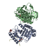


 Mass: 92.094 Da / Num. of mol.: 1 / Source method: obtained synthetically / Formula: C3H8O3 / Source: (synth.)
  Mass: 18.015 Da / Num. of mol.: 22 / Source method: isolated from a natural source / Formula: H2O
 Sample preparation
 Processing