 Movie
Movie Controller


+ Open data

- Basic information
| Entry | Database: PDB / ID: 6gxy | ||||||
|---|---|---|---|---|---|---|---|
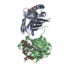
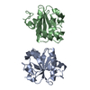
| Title | Tryparedoxin from Trypanosoma brucei in complex with CFT | ||||||
 Components Components | Tryparedoxin | ||||||
 Keywords Keywords |  OXIDOREDUCTASE / Covalent Inhibitor / Photoreduction / Inhibitor-Induced Dimerization / Monomer-Dimer Mixture OXIDOREDUCTASE / Covalent Inhibitor / Photoreduction / Inhibitor-Induced Dimerization / Monomer-Dimer Mixture | ||||||
| Function / homology |  Function and homology information Function and homology information | ||||||
| Biological species |  Trypanosoma brucei brucei (eukaryote) Trypanosoma brucei brucei (eukaryote) | ||||||
| Method | X-RAY DIFFRACTION / SYNCHROTRON / MOLECULAR REPLACEMENT / Resolution: 1.8 Å | ||||||
 Authors Authors | Bader, N. / Wagner, A. / Hellmich, U. / Schindelin, H. | ||||||
| Funding support |  Germany, 1items Germany, 1items
| ||||||
 Citation Citation | Journal: Angew. Chem. Int. Ed. Engl. / Year: 2019 Title: Inhibitor-Induced Dimerization of an Essential Oxidoreductase from African Trypanosomes. Authors: Wagner, A. / Le, T.A. / Brennich, M. / Klein, P. / Bader, N. / Diehl, E. / Paszek, D. / Weickhmann, A.K. / Dirdjaja, N. / Krauth-Siegel, R.L. / Engels, B. / Opatz, T. / Schindelin, H. / Hellmich, U.A. | ||||||
| History |
|
- Structure visualization
| Structure viewer | Molecule: MolmilJmol/JSmol |
|---|
- Downloads & links
-Download
| PDBx/mmCIF format | 6gxy.cif.gz | 118.3 KB | Display | PDBx/mmCIF format |
|---|---|---|---|---|
| PDB format | pdb6gxy.ent.gz | 91.5 KB | Display | PDB format |
| PDBx/mmJSON format | 6gxy.json.gz | Tree view | PDBx/mmJSON format | |
| Others |  Other downloads Other downloads |
-Validation report
| Arichive directory | https://data.pdbj.org/pub/pdb/validation_reports/gx/6gxyftp://data.pdbj.org/pub/pdb/validation_reports/gx/6gxy | HTTPS FTP |
|---|
-Related structure data
| Related structure data | 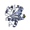 6gxgC 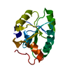 1o73S C: citing same article ( S: Starting model for refinement |
|---|---|
| Similar structure data |


-Links
 PDBj
PDBj

- Assembly
| Deposited unit | 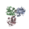
| ||||||||
|---|---|---|---|---|---|---|---|---|---|
| 1 | 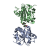
| ||||||||
| 2 | 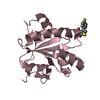
| ||||||||
| Unit cell |
|
-Components
| #1: Protein | Mass: 16019.042 Da / Num. of mol.: 3 Source method: isolated from a genetically manipulated source Source: (gene. exp.) Trypanosoma brucei brucei (eukaryote) / Production host:  Escherichia coli (E. coli) / References: UniProt: O77404 Escherichia coli (E. coli) / References: UniProt: O77404#2: Chemical |   Mass: 260.287 Da / Num. of mol.: 3 / Source method: obtained synthetically / Formula: C13H9FN2OS / Feature type: SUBJECT OF INVESTIGATION Mass: 260.287 Da / Num. of mol.: 3 / Source method: obtained synthetically / Formula: C13H9FN2OS / Feature type: SUBJECT OF INVESTIGATION#3: Water | ChemComp-HOH / | Water Mass: 18.015 Da / Num. of mol.: 624 / Source method: isolated from a natural source / Formula: H2O Mass: 18.015 Da / Num. of mol.: 624 / Source method: isolated from a natural source / Formula: H2O |
|---|
-Experimental details
-Experiment
| Experiment | Method: X-RAY DIFFRACTION / Number of used crystals: 1 |
|---|
- Sample preparation
| Crystal | Density Matthews: 1.87 Å3/Da / Density % sol: 34.3 % |
|---|---|
| Crystal grow | Temperature: 293 K / Method: vapor diffusion, sitting drop / pH: 5.5 / Details: 1.045 M Na-citrate, 0.1 M Na-acetate |
-Data collection
| Diffraction | Mean temperature: 100 K |
|---|---|
| Diffraction source | Source: SYNCHROTRON / Site: ESRF  / Beamline: ID23-1 / Wavelength: 0.980004 Å / Beamline: ID23-1 / Wavelength: 0.980004 Å |
| Detector | Type: DECTRIS PILATUS3 S 6M / Detector: PIXEL / Date: May 23, 2017 |
| Radiation | Protocol: SINGLE WAVELENGTH / Monochromatic (M) / Laue (L): M / Scattering type: x-ray |
| Radiation wavelength | Wavelength: 0.980004 Å / Relative weight: 1 |
| Reflection | Resolution: 1.8→50 Å / Num. obs: 32420 / % possible obs: 97.2 % / Redundancy: 4 % / CC1/2: 0.99 / Rmerge(I) obs: 0.141 / Rpim(I) all: 0.079 / Rrim(I) all: 0.163 / Net I/σ(I): 6.9 |
| Reflection shell | Resolution: 1.8→1.83 Å / Redundancy: 3.8 % / Rmerge(I) obs: 0.624 / Mean I/σ(I) obs: 1.8 / Num. unique obs: 1772 / CC1/2: 0.685 / Rpim(I) all: 0.354 / Rrim(I) all: 0.722 / % possible all: 90.2 |
- Processing
| Software |
| ||||||||||||||||||||||||||||||||||||||||||||||||||||||||||||||||||||||||||||||||||||||||||||||||||||||||||||||||||||||||||||||||||||||||||||||||||||||||||||||||||||||||||||||||||||||
|---|---|---|---|---|---|---|---|---|---|---|---|---|---|---|---|---|---|---|---|---|---|---|---|---|---|---|---|---|---|---|---|---|---|---|---|---|---|---|---|---|---|---|---|---|---|---|---|---|---|---|---|---|---|---|---|---|---|---|---|---|---|---|---|---|---|---|---|---|---|---|---|---|---|---|---|---|---|---|---|---|---|---|---|---|---|---|---|---|---|---|---|---|---|---|---|---|---|---|---|---|---|---|---|---|---|---|---|---|---|---|---|---|---|---|---|---|---|---|---|---|---|---|---|---|---|---|---|---|---|---|---|---|---|---|---|---|---|---|---|---|---|---|---|---|---|---|---|---|---|---|---|---|---|---|---|---|---|---|---|---|---|---|---|---|---|---|---|---|---|---|---|---|---|---|---|---|---|---|---|---|---|---|---|
| Refinement | Method to determine structure: MOLECULAR REPLACEMENT Starting model: 1o73 Resolution: 1.8→20 Å / Cor.coef. Fo:Fc: 0.961 / Cor.coef. Fo:Fc free: 0.93 / Cross valid method: THROUGHOUT / ESU R: 0.174 / ESU R Free: 0.147 / Details: HYDROGENS HAVE BEEN ADDED IN THE RIDING POSITIONS
| ||||||||||||||||||||||||||||||||||||||||||||||||||||||||||||||||||||||||||||||||||||||||||||||||||||||||||||||||||||||||||||||||||||||||||||||||||||||||||||||||||||||||||||||||||||||
| Solvent computation | Ion probe radii: 0.8 Å / Shrinkage radii: 0.8 Å / VDW probe radii: 1.2 Å | ||||||||||||||||||||||||||||||||||||||||||||||||||||||||||||||||||||||||||||||||||||||||||||||||||||||||||||||||||||||||||||||||||||||||||||||||||||||||||||||||||||||||||||||||||||||
| Displacement parameters | Biso mean: 20.198 Å2
| ||||||||||||||||||||||||||||||||||||||||||||||||||||||||||||||||||||||||||||||||||||||||||||||||||||||||||||||||||||||||||||||||||||||||||||||||||||||||||||||||||||||||||||||||||||||
| Refinement step | Cycle: 1 / Resolution: 1.8→20 Å
| ||||||||||||||||||||||||||||||||||||||||||||||||||||||||||||||||||||||||||||||||||||||||||||||||||||||||||||||||||||||||||||||||||||||||||||||||||||||||||||||||||||||||||||||||||||||
| Refine LS restraints |
|