 Movie
Movie Controller

[English] 日本語
 Yorodumi
- PDB-6gsu: FIRST-SPHERE AND SECOND-SPHERE ELECTROSTATIC EFFECTS IN THE ACTIV... -
+ Open data

- Basic information
| Entry | Database: PDB / ID: 6gsu | ||||||
|---|---|---|---|---|---|---|---|
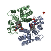
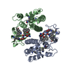
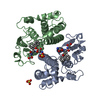
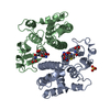
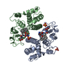
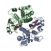
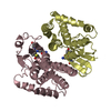
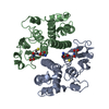
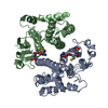
| Title | FIRST-SPHERE AND SECOND-SPHERE ELECTROSTATIC EFFECTS IN THE ACTIVE SITE OF A CLASS MU GLUTATHIONE TRANSFERASE | ||||||
 Components Components | MU CLASS GLUTATHIONE S-TRANSFERASE OF ISOENZYME 3-3 | ||||||
 Keywords Keywords |  TRANSFERASE / GLUTATHIONE TRANSFERASE / ISOENZYME 3-3 / T13A MUTANT TRANSFERASE / GLUTATHIONE TRANSFERASE / ISOENZYME 3-3 / T13A MUTANT | ||||||
| Function / homology |  Function and homology informationGlutathione conjugation / nitrobenzene metabolic process / cellular detoxification of nitrogen compound / hepoxilin biosynthetic process / glutathione binding / glutathione derivative biosynthetic process / response to metal ion / prostaglandin metabolic process / nickel cation binding / glutathione transferase ...Glutathione conjugation / nitrobenzene metabolic process / cellular detoxification of nitrogen compound / hepoxilin biosynthetic process / glutathione binding / glutathione derivative biosynthetic process / response to metal ion / prostaglandin metabolic process / nickel cation binding / glutathione transferase / glutathione transferase activity / response to axon injury / response to amino acid / glutathione metabolic process / xenobiotic catabolic process / steroid binding / response to lead ion / sensory perception of smell / cellular response to xenobiotic stimulus / response to ethanol / response to xenobiotic stimulus / protein kinase binding / enzyme binding / protein homodimerization activity / protein-containing complex / extracellular region / identical protein binding / cytosol / cytoplasm Function and homology informationGlutathione conjugation / nitrobenzene metabolic process / cellular detoxification of nitrogen compound / hepoxilin biosynthetic process / glutathione binding / glutathione derivative biosynthetic process / response to metal ion / prostaglandin metabolic process / nickel cation binding / glutathione transferase ...Glutathione conjugation / nitrobenzene metabolic process / cellular detoxification of nitrogen compound / hepoxilin biosynthetic process / glutathione binding / glutathione derivative biosynthetic process / response to metal ion / prostaglandin metabolic process / nickel cation binding / glutathione transferase / glutathione transferase activity / response to axon injury / response to amino acid / glutathione metabolic process / xenobiotic catabolic process / steroid binding / response to lead ion / sensory perception of smell / cellular response to xenobiotic stimulus / response to ethanol / response to xenobiotic stimulus / protein kinase binding / enzyme binding / protein homodimerization activity / protein-containing complex / extracellular region / identical protein binding / cytosol / cytoplasmSimilarity search - Function | ||||||
| Biological species |  Rattus rattus (black rat) Rattus rattus (black rat) | ||||||
| Method | X-RAY DIFFRACTION / Resolution: 1.85 Å | ||||||
 Authors Authors | Xiao, G. / Ji, X. / Armstrong, R.N. / Gilliland, G.L. | ||||||
 Citation Citation | Journal: Biochemistry / Year: 1996 Title: First-sphere and second-sphere electrostatic effects in the active site of a class mu gluthathione transferase. Authors: Xiao, G. / Liu, S. / Ji, X. / Johnson, W.W. / Chen, J. / Parsons, J.F. / Stevens, W.J. / Gilliland, G.L. / Armstrong, R.N. #1: Journal: Biochemistry / Year: 1994Title: Structure and Function of the Xenobiotic Substrate Binding Site of a Glutathione S-Transferase as Revealed by X-Ray Crystallographic Analysis of Product Complexes with the Diastereomers of 9- ...Title: Structure and Function of the Xenobiotic Substrate Binding Site of a Glutathione S-Transferase as Revealed by X-Ray Crystallographic Analysis of Product Complexes with the Diastereomers of 9-(S-Glutathionyl)-10-Hydroxy-9,10-Dihydrophenanthrene Authors: Ji, X. / Johnson, W.W. / Sesay, M.A. / Dickert, L. / Prasad, S.M. / Ammon, H.L. / Armstrong, R.N. / Gilliland, G.L. #2: Journal: J.Am.Chem.Soc. / Year: 1993Title: Second-Sphere Electrostatic Effects in the Active Site of Glutathione S-Transferase. Observation of an on-Facet Hydrogen Bond between the Side Chain of Threonine 13 and the Pi-Cloud of ...Title: Second-Sphere Electrostatic Effects in the Active Site of Glutathione S-Transferase. Observation of an on-Facet Hydrogen Bond between the Side Chain of Threonine 13 and the Pi-Cloud of Tyrosine 6 and its Influence on Catalysis Authors: Liu, S. / Ji, X. / Gilliland, G.L. / Stevens, W.J. / Armstrong, R.N. #3: Journal: Biochemistry / Year: 1993Title: Snapshots Along the Reaction Coordinate of an Snar Reaction Catalyzed by Glutathione Transferase Authors: Ji, X. / Armstrong, R.N. / Gilliland, G.L. #4: Journal: J.Biol.Chem. / Year: 1992Title: Contribution of Tyrosine 6 to the Catalytic Mechanism of Isoenzyme 3-3 of Glutathione S-Transferase Authors: Liu, S. / Zhang, P. / Ji, X. / Johnson, W.W. / Gilliland, G.L. / Armstrong, R.N. #5: Journal: Biochemistry / Year: 1992Title: The Three-Dimensional Structure of a Glutathione S-Transferase from the Mu Gene Class. Structural Analysis of the Binary Complex of Isoenzyme 3-3 and Glutathione at 2.2-A Resolution Authors: Ji, X. / Zhang, P. / Armstrong, R.N. / Gilliland, G.L. | ||||||
| History |
|
- Structure visualization
| Structure viewer | Molecule: MolmilJmol/JSmol |
|---|
- Downloads & links
-Download
| PDBx/mmCIF format | 6gsu.cif.gz | 112 KB | Display | PDBx/mmCIF format |
|---|---|---|---|---|
| PDB format | pdb6gsu.ent.gz | 86.1 KB | Display | PDB format |
| PDBx/mmJSON format | 6gsu.json.gz | Tree view | PDBx/mmJSON format | |
| Others |  Other downloads Other downloads |
-Validation report
| Arichive directory | https://data.pdbj.org/pub/pdb/validation_reports/gs/6gsuftp://data.pdbj.org/pub/pdb/validation_reports/gs/6gsu | HTTPS FTP |
|---|
-Related structure data


-Links
 PDBj
PDBj

- Assembly
| Deposited unit | 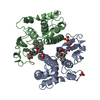
| |||||||||
|---|---|---|---|---|---|---|---|---|---|---|
| 1 |
| |||||||||
| Unit cell |
| |||||||||
| Components on special symmetry positions |
| |||||||||
| Noncrystallographic symmetry (NCS) | NCS oper: (Code: given Matrix: (0.819, -0.10421, 0.56425), Vector : |
-Components
| #1: Protein | Mass: 25788.764 Da / Num. of mol.: 2 / Mutation: T13A Source method: isolated from a genetically manipulated source Source: (gene. exp.) Rattus rattus (black rat) / Gene: CDNA INSERT OF CLONE PGT33MX / Organ: LIVER / Plasmid: PGT33MXT13A / Gene (production host): CDNA INSERT OF CLONE PGT33MX / Production host:  Escherichia coli (E. coli) / Strain (production host): M5219 / References: UniProt: P04905, glutathione transferase Escherichia coli (E. coli) / Strain (production host): M5219 / References: UniProt: P04905, glutathione transferase#2: Chemical | Sulfate  Mass: 96.063 Da / Num. of mol.: 3 / Source method: obtained synthetically / Formula: SO4 Mass: 96.063 Da / Num. of mol.: 3 / Source method: obtained synthetically / Formula: SO4#3: Chemical |   Mass: 501.552 Da / Num. of mol.: 2 / Source method: obtained synthetically / Formula: C24H27N3O7S Mass: 501.552 Da / Num. of mol.: 2 / Source method: obtained synthetically / Formula: C24H27N3O7S#4: Water | ChemComp-HOH / | Water Mass: 18.015 Da / Num. of mol.: 323 / Source method: isolated from a natural source / Formula: H2O Mass: 18.015 Da / Num. of mol.: 323 / Source method: isolated from a natural source / Formula: H2O |
|---|
-Experimental details
-Experiment
| Experiment | Method: X-RAY DIFFRACTION |
|---|
- Sample preparation
| Crystal | Density Matthews: 2.32 Å3/Da / Density % sol: 47 % | ||||||||||||||||||||||||||||||||||||||||
|---|---|---|---|---|---|---|---|---|---|---|---|---|---|---|---|---|---|---|---|---|---|---|---|---|---|---|---|---|---|---|---|---|---|---|---|---|---|---|---|---|---|
| Crystal grow | *PLUS pH: 8 / Method: vapor diffusion, sitting drop | ||||||||||||||||||||||||||||||||||||||||
| Components of the solutions | *PLUS
|
-Data collection
| Diffraction source | Wavelength: 1.5418 |
|---|---|
| Detector | Type: SIEMENS / Detector: AREA DETECTOR / Date: May 7, 1993 |
| Radiation | Monochromatic (M) / Laue (L): M / Scattering type: x-ray |
| Radiation wavelength | Wavelength: 1.5418 Å / Relative weight: 1 |
| Reflection | Resolution: 1.85→50 Å / Num. obs: 40404 / % possible obs: 95.6 % / Observed criterion σ(I): 0 / Redundancy: 3.55 % / Rmerge(I) obs: 0.09 |
| Reflection | *PLUS Num. obs: 38422 / Num. measured all: 149364 / Rmerge(I) obs: 0.059 |
- Processing
| Software |
| ||||||||||||||||||||||||||||||||||||||||||||||||||||||||||||||||||||||||||||||||||||
|---|---|---|---|---|---|---|---|---|---|---|---|---|---|---|---|---|---|---|---|---|---|---|---|---|---|---|---|---|---|---|---|---|---|---|---|---|---|---|---|---|---|---|---|---|---|---|---|---|---|---|---|---|---|---|---|---|---|---|---|---|---|---|---|---|---|---|---|---|---|---|---|---|---|---|---|---|---|---|---|---|---|---|---|---|---|
| Refinement | Resolution: 1.85→6 Å / σ(F): 4
| ||||||||||||||||||||||||||||||||||||||||||||||||||||||||||||||||||||||||||||||||||||
| Displacement parameters | Biso mean: 18.32 Å2 | ||||||||||||||||||||||||||||||||||||||||||||||||||||||||||||||||||||||||||||||||||||
| Refinement step | Cycle: LAST / Resolution: 1.85→6 Å
| ||||||||||||||||||||||||||||||||||||||||||||||||||||||||||||||||||||||||||||||||||||
| Refine LS restraints |
| ||||||||||||||||||||||||||||||||||||||||||||||||||||||||||||||||||||||||||||||||||||
| Software | *PLUS Name: GPRLSA / Classification: refinement | ||||||||||||||||||||||||||||||||||||||||||||||||||||||||||||||||||||||||||||||||||||
| Refinement | *PLUS Rfactor obs: 0.175 | ||||||||||||||||||||||||||||||||||||||||||||||||||||||||||||||||||||||||||||||||||||
| Solvent computation | *PLUS | ||||||||||||||||||||||||||||||||||||||||||||||||||||||||||||||||||||||||||||||||||||
| Displacement parameters | *PLUS |
