 Movie
Movie Controller

[English] 日本語
 Yorodumi
- PDB-6gmu: Serum paraoxonase-1 by directed evolution with the L69G/H134R/F22... -
+ Open data

- Basic information
| Entry | Database: PDB / ID: 6gmu | ||||||
|---|---|---|---|---|---|---|---|
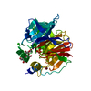
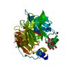
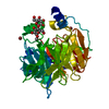
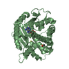
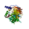
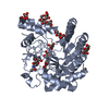
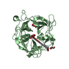
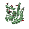
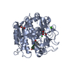
| Title | Serum paraoxonase-1 by directed evolution with the L69G/H134R/F222S/T332S mutations | ||||||
 Components Components | Serum Paraoxonase-1 by directed evolution with the L69G/H134R/F222S/T332S mutations | ||||||
 Keywords Keywords |  HYDROLASE / beta-propeller / Lactonase / HDL / Organophosphate-hydrolase HYDROLASE / beta-propeller / Lactonase / HDL / Organophosphate-hydrolase | ||||||
| Function / homology | TolB, C-terminal domain / 6 Propeller / Neuraminidase / Mainly Beta Function and homology information Function and homology information | ||||||
| Biological species |  Homo sapiens (human) Homo sapiens (human) | ||||||
| Method | X-RAY DIFFRACTION / SYNCHROTRON / MOLECULAR REPLACEMENT / Resolution: 2.7 Å | ||||||
 Authors Authors | Ben-David, M. / Sussman, J.L. / Tawfik, D.S. | ||||||
 Citation Citation | Journal: Mol.Biol.Evol. / Year: 2020 Title: Enzyme Evolution: An Epistatic Ratchet versus a Smooth Reversible Transition. Authors: Ben-David, M. / Soskine, M. / Dubovetskyi, A. / Cherukuri, K.P. / Dym, O. / Sussman, J.L. / Liao, Q. / Szeler, K. / Kamerlin, S.C.L. / Tawfik, D.S. | ||||||
| History |
|
- Structure visualization
| Structure viewer | Molecule: MolmilJmol/JSmol |
|---|
- Downloads & links
-Download
| PDBx/mmCIF format | 6gmu.cif.gz | 81.6 KB | Display | PDBx/mmCIF format |
|---|---|---|---|---|
| PDB format | pdb6gmu.ent.gz | 59.7 KB | Display | PDB format |
| PDBx/mmJSON format | 6gmu.json.gz | Tree view | PDBx/mmJSON format | |
| Others |  Other downloads Other downloads |
-Validation report
| Arichive directory | https://data.pdbj.org/pub/pdb/validation_reports/gm/6gmuftp://data.pdbj.org/pub/pdb/validation_reports/gm/6gmu | HTTPS FTP |
|---|
-Related structure data
| Related structure data |  6g82C  6h0aC 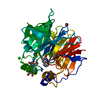 3srgS S: Starting model for refinement C: citing same article ( |
|---|---|
| Similar structure data |

-Links
 PDBj
PDBj
- Assembly
| Deposited unit | 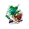
| ||||||||
|---|---|---|---|---|---|---|---|---|---|
| 1 |
| ||||||||
| Unit cell |
|
-Components
| #1: Protein | Mass: 39492.773 Da / Num. of mol.: 1 Source method: isolated from a genetically manipulated source Details: 1. There is no entry for this protein in the Uniprot, since the protein is a chimera (recombinant protein) of four genes (human, rabbit, mouse and rat). 2. The first 17 aa at the N-term is ...Details: 1. There is no entry for this protein in the Uniprot, since the protein is a chimera (recombinant protein) of four genes (human, rabbit, mouse and rat). 2. The first 17 aa at the N-term is missing due to lack of electron density. Source: (gene. exp.) Homo sapiens (human) / Production host:  Escherichia coli (E. coli) Escherichia coli (E. coli) | ||||||
|---|---|---|---|---|---|---|---|
| #2: Chemical |   Mass: 40.078 Da / Num. of mol.: 2 / Source method: obtained synthetically / Formula: Ca Mass: 40.078 Da / Num. of mol.: 2 / Source method: obtained synthetically / Formula: Ca#3: Chemical | Chloride  Mass: 35.453 Da / Num. of mol.: 3 / Source method: obtained synthetically / Formula: Cl Mass: 35.453 Da / Num. of mol.: 3 / Source method: obtained synthetically / Formula: Cl#4: Chemical | ChemComp-B3P / | Bis-tris propane  Mass: 282.334 Da / Num. of mol.: 1 / Source method: obtained synthetically / Formula: C11H26N2O6 / Comment: pH buffer*YM Mass: 282.334 Da / Num. of mol.: 1 / Source method: obtained synthetically / Formula: C11H26N2O6 / Comment: pH buffer*YM#5: Water | ChemComp-HOH / | Water Mass: 18.015 Da / Num. of mol.: 66 / Source method: isolated from a natural source / Formula: H2O Mass: 18.015 Da / Num. of mol.: 66 / Source method: isolated from a natural source / Formula: H2O |
-Experimental details
-Experiment
| Experiment | Method: X-RAY DIFFRACTION / Number of used crystals: 1 |
|---|
- Sample preparation
| Crystal | Density Matthews: 3.99 Å3/Da / Density % sol: 69.2 % |
|---|---|
| Crystal grow | Temperature: 291 K / Method: vapor diffusion, hanging drop Details: 0.1M Bis-Tris Propane pH 6.5, 0.2M Sodium Bromide and 20% PEG 3350 |
-Data collection
| Diffraction | Mean temperature: 100 K |
|---|---|
| Diffraction source | Source: SYNCHROTRON / Site: ESRF  / Beamline: ID23-1 / Wavelength: 0.9724 Å / Beamline: ID23-1 / Wavelength: 0.9724 Å |
| Detector | Type: ADSC QUANTUM 315 / Detector: CCD / Date: Nov 14, 2010 |
| Radiation | Protocol: SINGLE WAVELENGTH / Monochromatic (M) / Laue (L): M / Scattering type: x-ray |
| Radiation wavelength | Wavelength: 0.9724 Å / Relative weight: 1 |
| Reflection | Resolution: 2.7→50 Å / Num. obs: 18335 / % possible obs: 99.9 % / Redundancy: 13.5 % / Rmerge(I) obs: 0.157 / Rpim(I) all: 0.045 / Rrim(I) all: 0.163 / Net I/σ(I): 16.53 |
| Reflection shell | Resolution: 2.7→2.75 Å / Redundancy: 13.7 % / Rmerge(I) obs: 0.853 / Mean I/σ(I) obs: 2.96 / Num. unique obs: 881 / CC1/2: 0.927 / Rpim(I) all: 0.238 / Rrim(I) all: 0.886 / % possible all: 100 |
- Processing
| Software |
| ||||||||||||||||||||||||||||||||||||||||||||||||||||||||||||||||||||||||||||||||||||||||||||||||||
|---|---|---|---|---|---|---|---|---|---|---|---|---|---|---|---|---|---|---|---|---|---|---|---|---|---|---|---|---|---|---|---|---|---|---|---|---|---|---|---|---|---|---|---|---|---|---|---|---|---|---|---|---|---|---|---|---|---|---|---|---|---|---|---|---|---|---|---|---|---|---|---|---|---|---|---|---|---|---|---|---|---|---|---|---|---|---|---|---|---|---|---|---|---|---|---|---|---|---|---|
| Refinement | Method to determine structure: MOLECULAR REPLACEMENT Starting model: 3SRG Resolution: 2.7→33.308 Å / SU ML: 0.28 / Cross valid method: FREE R-VALUE / σ(F): 1.35 / Phase error: 21.38
| ||||||||||||||||||||||||||||||||||||||||||||||||||||||||||||||||||||||||||||||||||||||||||||||||||
| Solvent computation | Shrinkage radii: 0.9 Å / VDW probe radii: 1.11 Å | ||||||||||||||||||||||||||||||||||||||||||||||||||||||||||||||||||||||||||||||||||||||||||||||||||
| Refinement step | Cycle: LAST / Resolution: 2.7→33.308 Å
| ||||||||||||||||||||||||||||||||||||||||||||||||||||||||||||||||||||||||||||||||||||||||||||||||||
| Refine LS restraints |
| ||||||||||||||||||||||||||||||||||||||||||||||||||||||||||||||||||||||||||||||||||||||||||||||||||
| LS refinement shell |
|
