 Movie
Movie Controller


+ Open data

- Basic information
| Entry | Database: PDB / ID: 6gel | ||||||
|---|---|---|---|---|---|---|---|
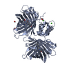
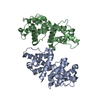
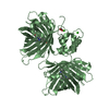
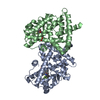
| Title | The structure of TWITCH-2B | ||||||
 Components Components | Green fluorescent protein,Optimized Ratiometric Calcium Sensor,Green fluorescent protein,Green fluorescent protein | ||||||
 Keywords Keywords |  FLUORESCENT PROTEIN / Twitch-2B / FRET / Ratiometric Biosensor FLUORESCENT PROTEIN / Twitch-2B / FRET / Ratiometric Biosensor | ||||||
| Function / homology |  Function and homology informationbioluminescence / generation of precursor metabolites and energy / calcium ion binding Function and homology informationbioluminescence / generation of precursor metabolites and energy / calcium ion bindingSimilarity search - Function | ||||||
| Biological species |   Aequorea victoria (jellyfish) Aequorea victoria (jellyfish) Opsanus tau (oyster toadfish) Opsanus tau (oyster toadfish) | ||||||
| Method | X-RAY DIFFRACTION / SYNCHROTRON / SAD / Resolution: 2.51 Å | ||||||
 Authors Authors | Trigo Mourino, P. / Paulat, M. / Thestrup, T. / Griesbeck, O. / Griesinger, C. / Becker, S. | ||||||
 Citation Citation | Journal: Sci Adv / Year: 2019 Title: Dynamic tuning of FRET in a green fluorescent protein biosensor. Authors: Trigo-Mourino, P. / Thestrup, T. / Griesbeck, O. / Griesinger, C. / Becker, S. #1: Journal: Nat. Methods / Year: 2014Title: Optimized ratiometric calcium sensors for functional in vivo imaging of neurons and T lymphocytes. Authors: Thestrup, T. / Litzlbauer, J. / Bartholomaus, I. / Mues, M. / Russo, L. / Dana, H. / Kovalchuk, Y. / Liang, Y. / Kalamakis, G. / Laukat, Y. / Becker, S. / Witte, G. / Geiger, A. / Allen, T. ...Authors: Thestrup, T. / Litzlbauer, J. / Bartholomaus, I. / Mues, M. / Russo, L. / Dana, H. / Kovalchuk, Y. / Liang, Y. / Kalamakis, G. / Laukat, Y. / Becker, S. / Witte, G. / Geiger, A. / Allen, T. / Rome, L.C. / Chen, T.W. / Kim, D.S. / Garaschuk, O. / Griesinger, C. / Griesbeck, O. | ||||||
| History |
|
- Structure visualization
| Structure viewer | Molecule: MolmilJmol/JSmol |
|---|
- Downloads & links
-Download
| PDBx/mmCIF format | 6gel.cif.gz | 439.5 KB | Display | PDBx/mmCIF format |
|---|---|---|---|---|
| PDB format | pdb6gel.ent.gz | 377.4 KB | Display | PDB format |
| PDBx/mmJSON format | 6gel.json.gz | Tree view | PDBx/mmJSON format | |
| Others |  Other downloads Other downloads |
-Validation report
| Arichive directory | https://data.pdbj.org/pub/pdb/validation_reports/ge/6gelftp://data.pdbj.org/pub/pdb/validation_reports/ge/6gel | HTTPS FTP |
|---|
-Related structure data
| Related structure data |  6gezC C: citing same article ( |
|---|---|
| Similar structure data | |
| Other databases |
|


-Links
 PDBj
PDBj

- Assembly
| Deposited unit | 
| |||||||||||||||||||||||||||||||||||||||||||||||||||||||||||||||||||||||||||||||||||||||||||||||||||||||||||||||||||||||||||||||||||||||||||||||||||||||||||||||||||||||||||||||||||||||||||||||||||||||||||||||||||||||||||
|---|---|---|---|---|---|---|---|---|---|---|---|---|---|---|---|---|---|---|---|---|---|---|---|---|---|---|---|---|---|---|---|---|---|---|---|---|---|---|---|---|---|---|---|---|---|---|---|---|---|---|---|---|---|---|---|---|---|---|---|---|---|---|---|---|---|---|---|---|---|---|---|---|---|---|---|---|---|---|---|---|---|---|---|---|---|---|---|---|---|---|---|---|---|---|---|---|---|---|---|---|---|---|---|---|---|---|---|---|---|---|---|---|---|---|---|---|---|---|---|---|---|---|---|---|---|---|---|---|---|---|---|---|---|---|---|---|---|---|---|---|---|---|---|---|---|---|---|---|---|---|---|---|---|---|---|---|---|---|---|---|---|---|---|---|---|---|---|---|---|---|---|---|---|---|---|---|---|---|---|---|---|---|---|---|---|---|---|---|---|---|---|---|---|---|---|---|---|---|---|---|---|---|---|---|---|---|---|---|---|---|---|---|---|---|---|---|---|---|---|---|
| 1 | 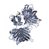
| |||||||||||||||||||||||||||||||||||||||||||||||||||||||||||||||||||||||||||||||||||||||||||||||||||||||||||||||||||||||||||||||||||||||||||||||||||||||||||||||||||||||||||||||||||||||||||||||||||||||||||||||||||||||||||
| 2 | 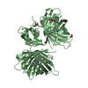
| |||||||||||||||||||||||||||||||||||||||||||||||||||||||||||||||||||||||||||||||||||||||||||||||||||||||||||||||||||||||||||||||||||||||||||||||||||||||||||||||||||||||||||||||||||||||||||||||||||||||||||||||||||||||||||
| Unit cell |
| |||||||||||||||||||||||||||||||||||||||||||||||||||||||||||||||||||||||||||||||||||||||||||||||||||||||||||||||||||||||||||||||||||||||||||||||||||||||||||||||||||||||||||||||||||||||||||||||||||||||||||||||||||||||||||
| Noncrystallographic symmetry (NCS) | NCS domain:
NCS domain segments:
NCS ensembles :
|
-Components
-Protein , 1 types, 2 molecules AB
| #1: Protein | Mass: 62449.891 Da / Num. of mol.: 2 Source method: isolated from a genetically manipulated source Source: (gene. exp.) Aequorea victoria (jellyfish), (gene. exp.) Opsanus tau (oyster toadfish)Gene: GFP / Production host:  Escherichia coli (E. coli) / Strain (production host): BL21(DE3) / References: UniProt: P42212, UniProt: W5IDB2 Escherichia coli (E. coli) / Strain (production host): BL21(DE3) / References: UniProt: P42212, UniProt: W5IDB2 |
|---|
-Non-polymers , 5 types, 127 molecules 


| #2: Chemical | ChemComp-CA /  Mass: 40.078 Da / Num. of mol.: 4 / Source method: obtained synthetically / Formula: Ca Mass: 40.078 Da / Num. of mol.: 4 / Source method: obtained synthetically / Formula: Ca#3: Chemical | Glycerol Mass: 92.094 Da / Num. of mol.: 3 / Source method: obtained synthetically / Formula: C3H8O3 Mass: 92.094 Da / Num. of mol.: 3 / Source method: obtained synthetically / Formula: C3H8O3#4: Chemical | Polyethylene glycol Mass: 194.226 Da / Num. of mol.: 2 / Source method: obtained synthetically / Formula: C8H18O5 / Comment: precipitant*YM Mass: 194.226 Da / Num. of mol.: 2 / Source method: obtained synthetically / Formula: C8H18O5 / Comment: precipitant*YM#5: Chemical | ChemComp-FMT / Formic acid Mass: 46.025 Da / Num. of mol.: 5 / Source method: obtained synthetically / Formula: CH2O2 Mass: 46.025 Da / Num. of mol.: 5 / Source method: obtained synthetically / Formula: CH2O2#6: Water | ChemComp-HOH / | WaterMass: 18.015 Da / Num. of mol.: 113 / Source method: isolated from a natural source / Formula: H2O |
|---|
-Experimental details
-Experiment
| Experiment | Method: X-RAY DIFFRACTION / Number of used crystals: 1 |
|---|
- Sample preparation
| Crystal | Density Matthews: 3.08 Å3/Da / Density % sol: 60.13 % |
|---|---|
| Crystal grow | Temperature: 293 K / Method: vapor diffusion / pH: 7 Details: 0.2 M sodium formiate, 5 mM calcium chloride, 18 % PEG 3350 |
-Data collection
| Diffraction | Mean temperature: 100 K |
|---|---|
| Diffraction source | Source: SYNCHROTRON / Site: SLS  / Beamline: X10SA / Wavelength: 1 Å / Beamline: X10SA / Wavelength: 1 Å |
| Detector | Type: DECTRIS PILATUS 6M / Detector: PIXEL / Date: Feb 24, 2014 |
| Radiation | Protocol: SINGLE WAVELENGTH / Monochromatic (M) / Laue (L): M / Scattering type: x-ray |
| Radiation wavelength | Wavelength: 1 Å / Relative weight: 1 |
| Reflection | Resolution: 2.51→47.93 Å / Num. obs: 50452 / % possible obs: 98.69 % / Redundancy: 6.62 % / Rrim(I) all: 0.033 / Net I/σ(I): 24.16 |
| Reflection shell | Resolution: 2.51→2.61 Å / Redundancy: 6.31 % / Mean I/σ(I) obs: 3.32 / Rrim(I) all: 0.241 |
- Processing
| Software |
| |||||||||||||||||||||||||||||||||||||||||||||||||||||||||||||||||||||||||||||||||||||||||||||||||||||||||||||||||||
|---|---|---|---|---|---|---|---|---|---|---|---|---|---|---|---|---|---|---|---|---|---|---|---|---|---|---|---|---|---|---|---|---|---|---|---|---|---|---|---|---|---|---|---|---|---|---|---|---|---|---|---|---|---|---|---|---|---|---|---|---|---|---|---|---|---|---|---|---|---|---|---|---|---|---|---|---|---|---|---|---|---|---|---|---|---|---|---|---|---|---|---|---|---|---|---|---|---|---|---|---|---|---|---|---|---|---|---|---|---|---|---|---|---|---|---|---|
| Refinement | Method to determine structure: SAD / Resolution: 2.51→47.93 Å / Cor.coef. Fo:Fc: 0.953 / Cor.coef. Fo:Fc free: 0.917 / SU B: 18.248 / SU ML: 0.196 / Cross valid method: THROUGHOUT / σ(F): 0 / ESU R: 0.372 / ESU R Free: 0.255 Details: HYDROGENS HAVE BEEN ADDED IN THE RIDING POSITIONS U VALUES : WITH TLS ADDED
| |||||||||||||||||||||||||||||||||||||||||||||||||||||||||||||||||||||||||||||||||||||||||||||||||||||||||||||||||||
| Solvent computation | Ion probe radii: 0.8 Å / Shrinkage radii: 0.8 Å / VDW probe radii: 1.2 Å | |||||||||||||||||||||||||||||||||||||||||||||||||||||||||||||||||||||||||||||||||||||||||||||||||||||||||||||||||||
| Displacement parameters | Biso max: 131.61 Å2 / Biso mean: 62.652 Å2 / Biso min: 23.29 Å2
| |||||||||||||||||||||||||||||||||||||||||||||||||||||||||||||||||||||||||||||||||||||||||||||||||||||||||||||||||||
| Refinement step | Cycle: final / Resolution: 2.51→47.93 Å
| |||||||||||||||||||||||||||||||||||||||||||||||||||||||||||||||||||||||||||||||||||||||||||||||||||||||||||||||||||
| Refine LS restraints |
| |||||||||||||||||||||||||||||||||||||||||||||||||||||||||||||||||||||||||||||||||||||||||||||||||||||||||||||||||||
| Refine LS restraints NCS | Refine-ID: X-RAY DIFFRACTION / Type: interatomic distance / Weight position: 0.05
| |||||||||||||||||||||||||||||||||||||||||||||||||||||||||||||||||||||||||||||||||||||||||||||||||||||||||||||||||||
| LS refinement shell | Resolution: 2.51→2.575 Å / Rfactor Rfree error: 0 / Total num. of bins used: 20
| |||||||||||||||||||||||||||||||||||||||||||||||||||||||||||||||||||||||||||||||||||||||||||||||||||||||||||||||||||
| Refinement TLS params. | Method: refined / Refine-ID: X-RAY DIFFRACTION
| |||||||||||||||||||||||||||||||||||||||||||||||||||||||||||||||||||||||||||||||||||||||||||||||||||||||||||||||||||
| Refinement TLS group |
|
